- Placental cotyledon
-
Placental cotyledon 
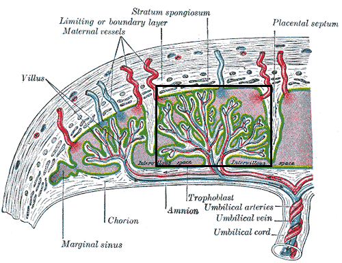
Structure of the placenta, with a placental cotyledon marked in rectangle. Gray's subject #12 63 In human development, the cotyledons are the approximately 15-30 separations of the desidua basalis of the placenta, separated by placental septa.[1] Each cotyledon consists of a main stem of a chorionic villus as well as its branches and subbranches etc.[1]
Vasculature
The cotyledons receive fetal blood from chorionic vessels, which branch off cotyledon vessels into the cotyledons, which, in turn, branch into capillaries.[2] The cotyledons are surrounded by maternal blood, which can exchange oxygen and nutrients with the fetal blood in the capillaries.
References
- ^ a b Page 565 in: Varney, Helen; Helen Varney Burst; Kriebs, Jan M.; Gegor, Carolyn L. (2004). Varney's midwifery. Boston: Jones and Bartlett Publishers. ISBN 0-7637-1856-4. [1]
- ^ Gordon, Z.; Elad, D.; Almog, R.; Hazan, Y.; Jaffa, A. J.; Eytan, O. (2007). "Anthropometry of fetal vasculature in the chorionic plate". Journal of Anatomy 211 (6): 698–706. doi:10.1111/j.1469-7580.2007.00819.x. PMC 2375851. PMID 17973911. http://www.pubmedcentral.nih.gov/articlerender.fcgi?tool=pmcentrez&artid=2375851.
External links

This anatomy article is a stub. You can help Wikipedia by expanding it.