- Circulating tumor cell
-
Circulating tumor cells (CTCs) are cells that have detached from a primary tumor and circulate in the bloodstream. CTCs may constitute seeds for subsequent growth of additional tumors (metastasis) in different tissues.
In 1869 Thomas Ashworth obeserved circulating tumor cells in the blood of a man with metastatic cancer. He postulated that “cells identical with those of the cancer itself being seen in the blood may tend to throw some light upon the mode of origin of multiple tumours existing in the same person”. A thorough comparison of the morphology of the circulating cells to tumor cells from different lesions led Ashworth to conclude that “One thing is certain, that if they [CTC] came from an existing cancer structure, they must have passed through the greater part of the circulatory system to have arrived at the internal saphena vein of the sound leg”.[1] Cancer research has demonstrated the critical role circulating tumor cells play in the metastatic spread of carcinomas.[2] Technology with the requisite sensitivity and reproducibility to detect CTC in patients with metastatic disease was developed only recently.[3][4][5][6][7]
Contents
Frequency of CTC
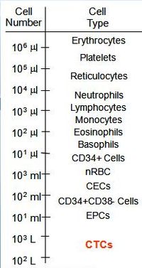 Figure 1: cell number of various blood cells in whole blood versus CTC
The detection of CTCs may have important prognostic and therapeutic implications but because their numbers can be very small, these cells are not easily detected.[8] Circulating tumor cells are found in frequencies in the order of 1-10 CTC per mL of whole blood in patients with metastatic disease. For comparison, a mL of blood contains a few million white blood cells and a billion red blood cells, see figure 1. This low frequency means that a key component of methods to detect CTC is the enrichment method.
First evidence indicates that CTC markers applied in human medicine are conserved in other species. Five of the more common markers including CK19 are also useful to detect CTC in the blood of dogs with malignant mammary tumors.[9]
Clinical utility
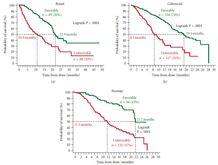
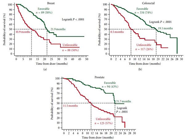 Figure 2: Kaplan Meier Analysis of overall survival before starting a new line of therapy for patients with metastatic breast, colorectal and prostate cancer. Patients were divided into those with Favorable and Unfavorable CTC (Unfavorable: >5 CTC/7.5mL for breast and prostate, >3 CTC/7.5mL for colon) [10]
To date, a variety of research methods have been developed to isolate and enumerate CTC.[11] The only U.S. Food and Drug Administration (FDA) cleared methodology for enumeration of CTC in whole blood is the CellSearch system.[12] Extensive clinical testing done using this method shows that presence of CTC is a strong prognostic factor for overall survival in patients with metastatic breast, colorectal or prostate cancer, see figure 2 [13][14][15][16][17][18][19]
CellSearch Method
Blood is sampled in an EDTA tube with an added preservative. Upon arrival in the lab, 7.5mL of blood is centrifuged and placed in a preparation system. This system first enriches the tumor cells immunomagnetically by means of ferrofluidic nanoparticles conjugated to epithelial cell adhesion molecule (EpCAM). Subsequently it stains the sample with a nuclear stain, and fluorescent antibody conjugates against CD45 and cytokeratin 8, 18 and 19 (CK). The sample is scanned on an analyzer, which takes images of the nuclear stain, the cytokeratin stain and the CD45 stain. A trained operator looks at the images of cells to identify those cells that are positive for the nuclear stain and the cytokeratin stain and negative for the CD45 stain. If the cell meets these criteria, the operator judges whether the morphology is suitable for a tumor cell. If the total number of tumor cells found is 5 or more, a sample is positive. In studies done on prostate, breast and colon cancer patients, median survival of metastatic patients with positive samples is about half the median survival of metastatic patients with negative samples.
Morphological definition
Morphological appearance is judged by human operators and is therefore subject to large inter operator variation.[20] Several CTC enumeration methods exist which use morphological appearance to identify CTC, which may also apply different morphological criteria. A recent study in prostate cancer showed that many different morphological definitions of circulating tumor cells have similar prognostic value, even though the absolute number of cells found in patients and normal donors varied by more than a decade between different morphological definitions.[21]
Further characterisation of CTC
Some drugs are particularly effective against cancers which fit certain requirements. For example Herceptin is very effective in patients who are Her2 positive, but much less effective in patients who are Her2 negative. Once the primary tumor is removed, biopsy of the current state of the cancer through traditional tissue typing is not possible anymore.[22] Often tissue sections of the primary tumor, removed years prior, are used to do the typing. Further characterisation of CTC may help determining the current tumor phenotype. FISH assays has been performed on CTC to as well as determination of IGF-1R, Her2, Bcl-2, [ERG (gene)|ERG], PTEN, AR status using immunofluorescence.[23][24][25][26][27][28]
See also
- Metastasis
- Tumor cell
References
- ^ Ashworth, T. R (1869). "A case of cancer in which cells similar to those in the tumours were seen in the blood after death". Australian Medical Journal 14: 146–7.
- ^ Fidler IJ (2003). "Timeline: The pathogenesis of cancer metastasis: the 'seed and soil' hypothesis revisited". Nat Rev Cancer 3 (6): 453–8. doi:10.1038/nrc1098. PMID 12778135.
- ^ Sleijfer S, Gratama JW, Sieuwerts AM, et al. (2007). "Circulating tumour cell detection on its way to routine diagnostic implementation?". Eur J Cancer 43 (18): 2645–50. doi:10.1016/j.ejca.2007.09.016. PMID 17977713.
- ^ Hayes DF, Smerage J. (2008). "Is There a Role for Circulating Tumor Cells in the Management of Breast Cancer?". Clin Cancer Res 14 (12): 3646–50. doi:10.1158/1078-0432.CCR-07-4481. PMID 18559576.
- ^ Pantel K, Alix-Panabières C, Riethdorf (2009). "Cancer micrometastases". Nat Rev Clin Oncol 6 (6): 339–51. doi:10.1038/nrclinonc.2009.44. PMID 19399023.
- ^ Pantel K, Riethdorf S. (2009). "Pathology: are circulating tumor cells predictive of overall survival?". Nat Rev Clin Oncol. 6 (4): 190–1. doi:10.1038/nrclinonc.2009.23. PMID 19333222.
- ^ Panteleakou Z, Lembessis P, Sourla A, et al. (2009). "Detection of circulating tumor cells in prostate cancer patients: methodological pitfalls and clinical relevance". Mol Med 15 (3-4): 101–14. doi:10.2119/molmed.2008.00116. PMC 2600498. PMID 19081770. http://www.pubmedcentral.nih.gov/articlerender.fcgi?tool=pmcentrez&artid=2600498.
- ^ Ghossein RA, Bhattacharya S, Rosai J (1999). "Molecular detection of micrometastases and circulating tumor cells in solid tumors". Clin. Cancer Res. 5 (8): 1950–60. PMID 10473071.
- ^ da Costa A, Oliveira JT, Gärtner F, Kohn B, Gruber AD, Klopfleisch R. (2010). "Potential markers for detection of circulating canine mammary tumor cells in the peripheral blood.". Veterinary Journal Epub Nov.2 2010. doi:10.1016/j.tvjl.2010.09.027. PMID 21051248.
- ^ MC Miller, GV Doyle, LWMM Terstappen (2010). "Significance of Circulating Tumor Cells detected by the CellSearch System in Patients with Metastatic Breast Colorectal and Prostate Cancer". Journal of Oncology 2010: 1. doi:10.1155/2010/617421.
- ^ Paterlini-Brechot P, Benali NL. (2007). "Circulating tumor cells (CTC) detection: Clinical impact and future directions". Cancer Lett. 253 (2): 180–204. doi:10.1016/j.canlet.2006.12.014. PMID 17314005.
- ^ "Veridex CellSearch Website". March 2010. http://veridex.com/CellSearch/CellSearchHCP.aspx. Retrieved 2010-03-14.
- ^ "Veridex LLC. CellSearch circulating tumor cell kit premarket notification—expanded indications for use—metastatic prostate cancer". March 2010. http://www.fda.gov/cdrh/pdf7/K073338.pdf. Retrieved 2010-03-14.[dead link]
- ^ Cristofanilli M, Budd GT, Ellis MJ, et al. (2004). "Circulating Tumor Cells, Disease Progression and Survival in Metastatic Breast Cancer". NEJM 351 (8): 781–91. doi:10.1056/NEJMoa040766. PMID 15317891.
- ^ Budd G, Cristofanilli M, Ellis M, et al. (2006). "Circulating Tumor Cells versus Imaging - Predicting Overall Survival in Metastatic Breast Cancer". Clin Can Res 12: 6404–09.
- ^ , et al. (2008). "The Relationship of Circulating Tumor Cells to Tumor Response, Progression-Free Survival, and Overall Survival in Patients with Metastatic Colorectal Cancer". JCO 26: 3213–21. doi:10.1200/JCO.2007.15.8923.
- ^ JS DeBono, HI Scher, RB Montgomery, et al. (2008). "Circulating Tumor Cells (CTC) predict survival benefit from treatment in metastatic castration resistant prostate cancer (CRPC)". Clin Can Res 14: 6302–9. doi:10.1158/1078-0432.CCR-08-0872.
- ^ Allard W J, Matera J, Miller MC, et al. (2004). "Tumor cells circulate in the peripheral blood of all major carcinomas but not in healthy subjects or patients with non-malignant diseases". Clin Can Res 10: 6897–6904. doi:10.1158/1078-0432.CCR-04-0378. PMID 15501967.
- ^ Riethdorf et al.; Fritsche, H; Müller, V; Rau, T; Schindlbeck, C; Rack, B; Janni, W; Coith, C et al. (2007). "Detection of Circulating Tumor Cells in Peripheral Blood of Patients with Metastatic Breast Cancer: A Validation Study of the CellSearch System". Clin Cancer Res 13 (3): 920–8. doi:10.1158/1078-0432.CCR-06-1695. PMID 17289886.
- ^ AGJ Tibbe, MC Miller, LWMM Terstappen (2007). "Statistical Considerations for Enumeration of Circulating Tumor Cells". Cytometry Part A 71A: 132–142.
- ^ F. A. W. Coumans, C. J. M. Doggen, G. Attard, et al. (2010). "All circulating EpCAM1CK1CD452 objects predict overall survival in castration-resistant prostate cancer". Ann of Oncology 21: 1851. doi:10.1093/annonc/mdq030.
- ^ Meng S, Tripathy D, Shete S et al. (2004). "HER-2 Gene Amplification can be acquired as breast cancer progresses". PNAS 101 (25): 9393–8. doi:10.1073/pnas.0402993101. PMC 438987. PMID 15194824. http://www.pubmedcentral.nih.gov/articlerender.fcgi?tool=pmcentrez&artid=438987.
- ^ Hayes DF, Walker TM, Singh B, et al. (2002). "Monitoring Expression of HER-2 on Circulating Epithelial Cells in Patients with advanced Breast Cancer". Int J of Oncology 21: 1111–8.
- ^ O'Hara SM, Moreno JG, Zweitzig DR, et al. (2004). "Multigene Reverse Transcription-PCR Profiling of Circulating Tumor Cells in Hormone-Refractory Prostate Cancer". Clin Chem 50 (5): 826–835. doi:10.1373/clinchem.2003.028563. PMID 14988224.
- ^ de Bono JS, Attard G, Adjei A, et al. (2007). "Potential Applications for Circulating Tumor Cells expressing the Insulin Growth Factor-I Receptor". Clin Can Res 13: 3611–6. doi:10.1158/1078-0432.CCR-07-0268.
- ^ Attard G, Swennenhuis JF, Olmos D et al. (2009). "Characterization of ERG, AR and PTEN status in Circulating Tumor Cells from Patients with Castration-Resistant Prostate Cancer". Cancer Research 69 (7): 2912–8. doi:10.1158/0008-5472.CAN-08-3667. PMID 19339269.
- ^ Swennenhuis JF, Tibbe AGJ, Levink R et al. (2009). "Characterization of Circulating Tumor Cells by Fluorescence In-Situ Hybridization". Cytometry Part A 75A: 520–7. doi:10.1002/cyto.a.20718.
- ^ Karp DD, Pollak MN, Cohen RB et al. (2009). "Pharmacokinetics and Pharmacodynamics of the IGF-IR Inhibitor Figitumumab (CP-751,871) in Combination with Paclitaxel and Carboplatin". Journal of Thoracic Oncology 4 (11): 1397–1403. doi:10.1097/JTO.0b013e3181ba2f1d. PMC 2941876. PMID 19745765. http://www.pubmedcentral.nih.gov/articlerender.fcgi?tool=pmcentrez&artid=2941876.
Categories:

Wikimedia Foundation. 2010.