- Superior oblique myokymia
Infobox_Disease
Name = Superior oblique myokymia
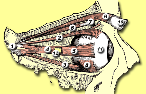
Caption = 6 =Superior oblique muscle
DiseasesDB =
ICD10 =
ICD9 =
ICDO =
OMIM =
MedlinePlus =
eMedicineSubj =
eMedicineTopic =
MeshID =Superior oblique myokymia is a
neurological disorder affecting vision and was termed by Hoyt and Keane in 1970. [cite journal |author=Hoyt WF, Keane JR. |title=Superior oblique myokymia: report and discussion on five case of benign intermittent uniocular microtremor. |journal=Arch Ophthalmol |year=1970 |volume=84 |pages=461–7]It is a condition that presents as repeated, brief episodes of movement, shimmering or shaking of the vision of one eye, a feeling of the eye trembling, or vertical/tilted vision. It can present as one or more of these symptoms. Diagnosis is most often made by the elimination of other conditions, disorders or diseases.
Onset usually occurs in adulthood, and the course is benign and is not commonly associated with other disorders.
Causes
In 1983, Bringewald postulated that superior oblique myokymia resulted from
vascular compression of thetrochlear nerve (fourth cranial nerve), which controls the action of thesuperior oblique muscle in the eye. [cite journal |author=Bringewald PR |title=Superior oblique myokymia |journal=Arch Neurol |year=1983 |volume=40 |pages=526] By 1998, there had been only one reported case of compression of the trochlear nerve by vessels. [cite journal |author=Samii M, Rosahl SK, Carvalho GA, Krzizok T |title=Microvascular decompression for superior oblique myokymia: first experience. Case report |journal=J. Neurosurg. |volume=89 |issue=6 |pages=1020–4 |year=1998 |pmid=9833830 |doi=] [cite journal |author=Scharwey K, Krzizok T, Samii M, Rosahl SK, Kaufmann H |title=Remission of superior oblique myokymia after microvascular decompression |journal=Ophthalmologica |volume=214 |issue=6 |pages=426–8 |year=2000 |pmid=11054004 |doi= ] More recently,magnetic resonance imaging experiments have shown that neurovascular compression at the root exit zone of the trochlear nerve can result in superior oblique myokymia. [cite web |url=http://www.ncbi.nlm.nih.gov/sites/entrez?db=PubMed&cmd=Retrieve&list_uids=11891831&dopt=Abstract |title=Superior oblique myokymia: magnetic resonance imaging support for the neurovascular compression hypothesis PMID: 11891831 |accessdate=2007-06-26 |format= |work= ]Treatment
Treatment can include pharmaceutical or surgical means. The drug carbamazepine (Tegretol) has been used successfully. Other drugs used with variable success include gabapentin and, recently, memantine. Successful surgery options include superior oblique tenectomy accompanied by inferior oblique myectomy. [cite web |url=http://telemedicine.orbis.org/bins/volume_page.asp?cid=735-954-1328-1350&lang=1 |title=Superior Oblique Myokymia 379.58 |accessdate=2007-06-25 |format= |work=]
Samii et al [cite journal |author=Samii M, Rosahl SK, Carvalho GA, Krzizok T |title=Microvascular decompression for superior oblique myokymia: first experience. Case report |journal=J. Neurosurg. |volume=89 |issue=6 |pages=1020–4 |year=1998 |pmid=9833830 |doi=] and Scharwey and Samii [cite journal |author=Scharwey K, Krzizok T, Samii M, Rosahl SK, Kaufmann H |title=Remission of superior oblique myokymia after microvascular decompression |journal=Ophthalmologica |volume=214 |issue=6 |pages=426–8 |year=2000 |pmid=11054004 |doi=] described a patient who had superior oblique myokymia for 17 years. The interposition of a
Teflon pad between thetrochlear nerve and a compressing artery and vein at the nerve's exit from the midbrain led to a remission lasting for a follow-up of 22 months.References
Other Resources
* [http://groups.msn.com/Superiorobliquemyokymia/somqapage1.msnw SOMPeople FAQ page]
Wikimedia Foundation. 2010.