- Lacuna (histology)
Infobox Anatomy
Name = Lacuna (histology)
Latin =
GraySubject = 18
GrayPage = 90
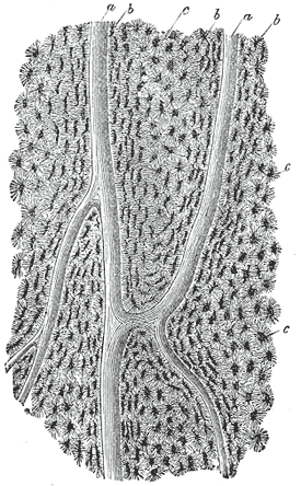
Caption = Section parallel to the surface from the body of thefemur . X 100. a,Haversian canals ; b, lacunae seen from the side; c, others seen from the surface inlamellae , which are cut horizontally.
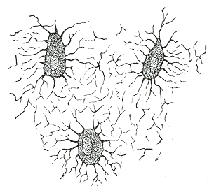
Caption2 = Nucleated bone cells and their processes, contained in the bone lacunæ and theircanaliculi respectively. From a section through thevertebra of an adultmouse .
System =
MeshName =
MeshNumber =
DorlandsPre = l_01
DorlandsSuf = 12475145
Inhistology , a lacuna is a small space containing anosteocyte in bone orchondrocyte in cartilage.Bone
The Lacunæ are situated between the lamellæ, and consist of a number of oblong spaces. In an ordinary microscopic section, viewed by transmitted light, they appear as fusiform opaque spots. Each lacuna is occupied during life by a branched cell, termed an
osteocyte , bone-cell or bone-corpuscle. Lacunae are connected to one another by small canals calledcanaliculi .Cartilage
The cells are contained in cavities in the matrix, called cartilage lacunæ; around these the matrix is arranged in concentric lines, as if it had been formed in successive portions around the cartilage cells. This constitutes the so-called capsule of the space. Each lacuna is generally occupied by a single cell, but during the division of the cells it may contain two, four, or eight cells. Lacunae are found between narrow sheets of calcified matrix that are known as lamellae (lah-MEL-le).
External links
*
* [http://www.mansfield.ohio-state.edu/~jbradley/BoneModelA.html Photo at ohio-state.edu]
Wikimedia Foundation. 2010.