- Mitral valve annuloplasty
-
Mitral valve annuloplasty is a surgical technique for the repair of leaking mitral valves. Due to various factors, the two leaflets normally involved in sealing the mitral valve to reterograde flow, may not coapt properly. Surgical repair typically involves the implantation of a device surrounding the mitral valve, called an annuloplasty device, which pulls the leaflets together to facilitate coaptation and aids to re-establish mitral valve function.
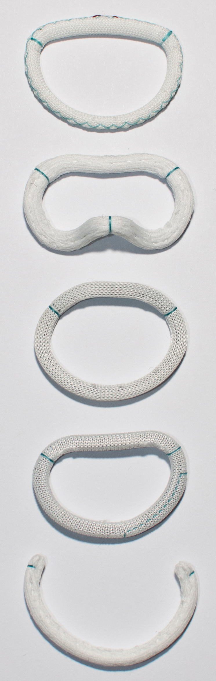
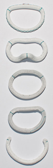 Examples for commercially available annuloplasty rings. From top: St. Judes Rigid Saddle Shaped Ring, Edwards Geoform, Edwards Physio, Edwards ETlogix, Edwards Cosgrove.
Contents
Need
Mitral regurgitation is the most common form of mitral valve dysfunction. Today more than 2.5 million Americans are estimated to be affected by mitral regurgitation. This number is expected to double by the year 2030. Every year, 300,000 people worldwide undergo open heart surgery for mitral valve repair, 44,000 people in the US alone.[1]
Relevance
Since it was initially established 40 years ago by Professor Alain Carpentier, mitral valve annuloplasty has been continuously improved and is considered the gold standard for the treatment of most etiologies of mitral valve dysfunction today.[2]
Rationale
The goal of mitral valve annuloplasty is to regain mitral valve competence by restoring the physiological form and function of the normal mitral valve apparatus.[3][4] Under normal conditions the mitral valve undergoes significant dynamic changes in shape and size throughout the cardiac cycle.[5] These changes are primarily due to the dynamic motion of the surrounding mitral valve annulus,[6][7] a collageneous structure which attaches the mitral leaflets and the left atrium to the ostium of the left ventricle and the aortic root.[8] Throughout the cardiac cycle, the annulus undergoes a sphincter motion, narrowing down the orifice area during systole to facilitate coaptation of the two leaflets and widens during diastole to allow for easy diastolic filling of the left ventricle. This motion is further enhanced by a pronounced three-dimensional configuration during systole, the characteristic saddle shape.[9][10] These changes throughout the cycle are believed to be key to optimize leaflet coaptation [11] and to minimize tissue stresses.[12] The challenge of mitral valve annuloplasty is to improve the diseased and distorted shape of the mitral valve, and to reestablish the physiological configuration, while preserving normal annular dynamics.[13] Today, cardiac surgeons can select from a variety of annuloplasty devices.[14] Devices are different in that they can be flexible, semi-rigid, or rigid, incomplete or complete, planar or saddle-shaped, adjustable and non-adustable. While the general goal of all devices is the same, namely to increase leaflet coaptation and to support the posterior annulus against dilation, flexible bands are designed to maintain the three-dimensional contour of the native annulus and some of its natural dynamics.[15] The goal of semi-rigid ring is to maintain coaptation and valve integrity during systole, while allowing for good hemodynamics during diastole.[16] Rigid rings are designed to provide rigid support in large dilation and under high-pressure.[17]
Sizing
Annuloplasty devices usually come in different sizes. The surgeon can estimate the dimensions of the patient’s mitral valve and decide on an appropriate ring size. Depending on the disease etiology different approaches are taken. Surgeons can decide to “undersize” the device, “truesize” the device, or “oversize”.
Device classification and regulatory considerations
Annuloplasty devices are classified by the U.S. Food and Drug Administration (FDA) as class II medical device. According to the FDA following issues need to be addressed before annuloplasty rings can be approved for marketing [18]:
Performance testing
- Biocompatibility testing
- Computational structural analysis
- Tensile testing
- Suture pull-out testing
- Sterilization validation
- Biological testing including bioburden and pyrogen testing
- Shelf-life validation
Labeling
- Contraindications
- Potential complications
- Instructions for use
- Specific warnings of potential risks
Commercially available devices
Edwards Life Science
- Carpentier-Edwards Classic Annuloplasty Ring
- Carpentier-Edwards Physio Annuloplasty Ring
- Carpentier-Edwards Physio II Annuloplasty Ring
- Carpentier-McCarthy-Adams IMR ETlogix Annuloplasty Ring
- GeoForm Ring
- Cosgrove-Edwards Annuloplasty System
St. Jude Medical
- St. Jude Medical Séguin Annuloplasty Ring
- St. Jude Medical Tailor Annuloplasty Ring
- St. Jude Medical Tailor Annuloplasty Band
- St. Jude Medical Rigid Saddle Ring
- St. Jude Medical Attune Annuloplasty Ring
Medtronic
- Profile 3D Ring
- CG Future Ring and Band System
- Duran AnCore Ring and Band System
- Simulus Adjustable Annuloplasty System
- Simulus Flexible Annuloplasty System
- Simulus Semi-Rigid Annuloplasty System
Minimally invasive annuloplasty
New technologies that allow for minimally invasive surgery have also begun to change mitral valve surgery. A number of devices and techniques have been developed that do not necessitate open-heart surgery for the repair of the mitral valve. Following is a list of minimally invasive annuloplasty devices or related [19]:
Coronary Sinus Delivery
- Cardiac Dimensions
- Mitralign
- Monarc (Edwards LifeSciences)
- Viaco
Other
- Accucinch
- ExTensia
- MitralSolution
References
- ^ Bonow, R.O.; et al. (2006). "ACC/AHA 2006 guidelines for the management of patients with valvular heart disease". Circulation 114: e84-e231.
- ^ de Oliveira, J.M.F; Antunes, M.J (2006). "Mitral valve repair: better than replacement". Heart 92: 275–281.
- ^ Carpentier, A.F. (2010). Carpentier’s Reconstructive Valve Surgery. Elsevier Saunders.
- ^ Rausch, M.K.; Bothe W., Kvitting J.P., Swanson J.C., Miller D.C., Kuhl E. (2011). "Mitral valve annuloplasty - A quantitative clinical and mechanical comparison of different annuloplasty devices". Annals of Biomedical Engineering. doi:10.1007/s10439-011-0442-y.
- ^ Ormiston, J.A.; Shah, P.M., Tei, C., Wong, M (1981). "Size and motion of the mitral valve annulus in man. I. A twodimensional echocardiographic method and findings in normal subjects". Circulation 64: 113–120.
- ^ Rausch, M.K.; Bothe, W., Kvitting, J.P.E., Goktepe, S., Miller, D.C., Kuhl, E (2011). "In vivo dynamic strains of the ovine anterior mitral valve leaflet". Journal of Biomechanics 44: 1149–1157.
- ^ Rausch, M.K.; Bothe, W., Kvitting, J.P.E., Swanson, J.C., Ingels, N.B., Miller, D.C., Kuhl, E (2011). "Characterization of mitral valve annular dynamics in the beating heart". Annals of Biomedical Engineering 39: 1690–1702.
- ^ Kaplan, S.R.; Bashein, G., Sheehan, F.H., Legget, M.E., Munt, B., Li, X.N., Sivarajan, M., Bolson, E.L., Zeppa, M., Arch, M.Z., Martin, R.W (2000). "Three-dimensional echocardiographic assessment of annular shape changes in the normal and regurgitant mitral valve". American Heart Journal 139: 378–387.
- ^ Jimenez, J.H.; Soerensen, D.D., He, Z., He, S., Yoganathan, A.P (2003). "Effects of a saddle shaped annulus on mitral valve function and chordal force distribution: an in vitro study". Annals of Biomedical Engineering 31: 1171–1181.
- ^ Levine, R.A.; Handschumacher, M.D., Sanfilippo, A.J., Hagege, A.A., Harrigan, P., Marshall, J.E., Weyman, A.E (1980). "Three-dimensional echocardiographic reconstruction of the mitral valve, with implications for the diagnosis of mitral valve prolapse". Circulation 89: 589–598.
- ^ Jensen, M.O.; Jensen, H., Smerup, M., Levine, R.A., Yoganathan, A.P., Nygaard, H., Hasenkam, J.M., Nielsen, S.L. (2008). "Saddle-shaped mitral valve annuloplasty rings experience lower forces compared with flat rings". Circulation 118: 250–255.
- ^ Padala, M; Hutchison, R.A., Croft, L.R., Jimenez, J.H., Gorman, R.C., Gorman, J.H., Sacks, M.S., Yoganathan, A.P. (2009). "Saddle shape of the mitral annulus reduces systolic strains on the P2 segment of the posterior mitral leaflet". Annals of Thoracic Surgery 88: 1499–1504.
- ^ Cosgrove, D.M.; Arcidi, J.M., Rodriguez, L., Stewart, W.J., Powell, K., Thomas, J.D. (1995). "Initial experience with the Cosgrove-Edwards Annuloplasty System". Annals of Thoracic Surgery 60: 499–503.
- ^ Bothe, W.; Kuhl, E., Kvitting, J.P.E., Rausch, M.K., Goktepe, S., Swanson, J., S, F., Ingels, N., Miller, D. (2011). "Rigid, complete annuloplasty rings increase anterior mitral leaflet strains in the normal beating ovine heart". Circulation 124: 81–96.
- ^ Cosgrove, D.M.; Arcidi, J.M., Rodriguez, L., Stewart, W.J., Powell, K., Thomas, J.D. (1995). "Initial experience with the Cosgrove-Edwards Annuloplasty System". Annals of Thoracic Surgery 60: 499–503.
- ^ Carpentier, A.F.; Lessana, A., Relland, J.Y., Belli, E., Mihaileanu, S., Berrebi, A.J., Palsky, E., Loulmet, D.F. (1995). "The"Physio-Ring": an advanced concept in mitral valve annuloplasty". Annals of Thoracic Surgery 60: 1177–1186.
- ^ Votta, E.; Maisano, F., Bolling, S.F., Alfieri, O., Montevecchi, F.M., Redaelli, A (2007). "The Geoform disease-specific annuoloplasty system: a finite element study". Annals of Thoracic Surgery 84: 92–102.
- ^ "Guidance for Annuloplasty Rings 510(k) Submissions; Final Guidance for Industry and FDA Staff". FDA. http://www.fda.gov/downloads/MedicalDevices/DeviceRegulationandGuidance/GuidanceDocuments/ucm073648.pdf. Retrieved 15 November 2011.
- ^ Mack, M.J. (2006). "New techniques for percutaneous repair of the mitral valve". Heart Failure Review 11: 259–268.
Wikimedia Foundation. 2010.