endocardial cushions — elevations of embryonic connective tissue covered by endothelium bulging into the atrioventricular canal of the embryonic heart, which later fuse with the free edge of the septum primum to separate the right and left atria … Medical dictionary
endocardial cushion defects — a spectrum of septal defects resulting from imperfect fusion of the endocardial cushions and ranging from persistent ostium primum to persistent complete common atrioventricular canal; see atrial septal d s and atrioventricularis communis … Medical dictionary
cushion — In anatomy, any structure resembling a pad or c.. anal cushions vascular prominences formed by clusters of normally sacculated veins of the superior rectal venous plexus, fed by arteriovenous anastomoses that cause their … Medical dictionary
Congenital heart defect — Classification and external resources The normal structure of the heart (left) in comparison to two common locations for a ventricular septal defect (right), the most common form of congenital heart defect.[1] … Wikipedia
human cardiovascular system — ▪ anatomy Introduction organ system that conveys blood through vessels to and from all parts of the body, carrying nutrients and oxygen to tissues and removing carbon dioxide and other wastes. It is a closed tubular system in which the… … Universalium
Septum — A word borrowed from the Latin "saeptum" meaning a "dividing wall or enclosure." * * * 1. [TA] A thin wall dividing two cavities or masses of softer tissue. See septal area, transparent s.. 2. In fungi, a wall; usually a cross … Medical dictionary
Atrioventricular septal defect — Classification and external resources ICD 10 Q21.2 ICD 9 745.6 … Wikipedia
Truncus arteriosus (embryology) — Infobox Embryology Name = Truncus arteriosus Latin = GraySubject = 135 GrayPage = 514 Caption = Heart of human embryo of about fourteen days. (Truncus arteriosis visible at top.) Caption2 = Diagrams to illustrate the transformation of the bulbus… … Wikipedia
Aortic septum — Infobox Embryology Name = PAGENAME Latin = GraySubject = 135 GrayPage = 514 Caption = Heart of human embryo of about thirty five days, opened on right side. (Aortic septum labeled at center right.) Caption2 = Diagrams to show the development of… … Wikipedia
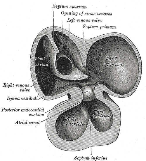
Septum intermedium — Infobox Embryology Name = PAGENAME Latin = GraySubject = 135 GrayPage = 512 Caption = Interior of dorsal half of heart of human embryo of about thirty five days. Caption2 = Same heart as in Fig. 467, opened on right side. Days = 35 System =… … Wikipedia