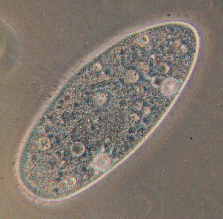
- Peniculid
Taxobox | color = #FA7B62
name = Peniculids

image_caption = "Paramecium aurelia"
domain = Eukaryota
regnum =Chromalveolata
superphylum =Alveolata
phylum = Ciliophora
classis =Oligohymenophorea
ordo = Peniculida
ordo_authority =Fauré-Fremiet inCorliss 1956
subdivision_ranks = Typical families
subdivision = SuborderFrontoniina
Clathrostomatidae
Frontoniidae
Lembadionidae
Maritujidae
Stokesiidae
SuborderParameciina
Parameciidae
Neobursaridiidae
Urocentridae The peniculids are an order ofciliate protozoa, including the well-known "Paramecium " and its close relatives. Most are relatively large, freshwater forms that feed on smaller organisms swept into the mouth. They have simple life cycles, and in many cases do not even form resting cysts.Typically the body has uniform, dense cilia, which also cover a vestibule preceding the mouth.
Extrusome s are characteristically in the form of spindle trichocysts, which release thread-like shafts, and never mucocysts. The oral cilia include peniculi, corresponding to the membranelles of related groups, arranged parallel to the mouth deep in the oral cavity. Nematodesmata (rods) arise from the bases of the oral or perioral cilia, but these do not support a cyrtos as in some other classes. Two suborders are recognised:* The Frontoniina typically have a shallow oral cavity, with a long paroral membrane and denser somatic kineties to the right of the mouth. These are called "ophryokineties", and take part in forming the new mouth during cell division.
* The Parameciina typically have a deeper oral cavity, with peniculi mainly forward of the mouth, and the paroral membrane reduced, though still present throughout interphase.
The peniculids were first defined by
Fauré-Fremiet in 1956. Originally they were one of three suborders of thehymenostome s, which are now treated as subclasses of theOligohymenophorea . They were divided into two suborders by Small and Lynn in 1985, who placed them in the classNassophorea , owing to ultrastructural peculiarities such as the presence of nematodesmata, which was considered to indicate the cyrtos was secondarily absent. However, more recent schemes reverse this move.
Wikimedia Foundation. 2010.
