Ossification of ulna

Ossification of ulna
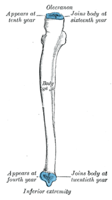
Plan of ossification of the ulna. From three centers.
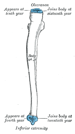
Epiphysial lines of ulna in a young adult. Lateral aspect. The lines of attachment of the articular capsules are in blue.

The ulna is ossified from three centers: one each for the body, the inferior extremity, and the top of the olecranon.

Ossification begins near the middle of the body, about the eighth week of fetal life, and soon extends through the greater part of the bone.

At birth the ends are cartilaginous.

About the fourth year, a center appears in the middle of the head, and soon extends into the styloid process.

About the tenth year, a center appears in the olecranon near its extremity, the chief part of this process being formed by an upward extension of the body.

The upper epiphysis joins the body about the sixteenth, the lower about the twentieth year.


Wikimedia Foundation. 2010.

Игры ⚽ Нужна курсовая?

Look at other dictionaries:

  • Ossification center — Section of fetal bone of cat. ir. Irruption of the subperiosteal tissue. p. Fibrous layer of the periosteum. o. Layer of osteoblasts. im. Subperiosteal bony deposit. Latin centrum ossificationis Gray …   Wikipedia

  • Ossification — Bone is broken down by osteoclasts, and rebuilt by osteoblasts, both of which communicate through cytokine (TGF β, IGF) signalling. Ossification (or osteogenesis) is the process of laying down new bone material by cells called osteoblasts. It is… …   Wikipedia

  • Ossification of scapula — Figure 5 : Plan of ossification of the scapula. From seven centers. The scapula is ossified from 7 or more centers: one for the body, two for the coracoid process, two for the acromion, one for the vertebral border, and one for the inferior… …   Wikipedia

  • Ossification of humerus — Figure 4 : Plan of ossification of the humerus. Figure 5 : Epiphysial lines of humerus in a young adult. Anterior aspect. The lines of attachment of the articular …   Wikipedia

  • Ossification of tibia — Plan of ossification of the tibia. From three centers. Epiphysial lines of tibia and fibula in a young adult. Anter …   Wikipedia

  • Ossification of radius — Figure 1 : Plan of ossification of the radius. From three centers. Figure 2 : Epiphysial lines of radius in a young adult. Anterior aspect. The line of attachmen …   Wikipedia

  • Ossification of mandible — Figure 3: Mandible of human embryo 24 mm. long. Outer aspect. Figure 4: Mandible of human embryo 24 mm. long. Inner …   Wikipedia

  • Ossification of ethmoid — Bone: Ossification of ethmoid Sagittal section of skull. (Ethmoid bone visible as white structure to left.) Gray s subject …   Wikipedia

  • Ossification of temporal bone — 1. Outer surface of petromastoid part. 2. Outer surface of tympanic ring. 3. Inner surface of squama. Figure 7& …   Wikipedia

  • Ossification of occipital bone — Figure 3 : Occipital bone at birth. The planum occipitale [Fig. 3] of the squama of the occipital bone is developed in membrane, and may remain separate throughout life when it constitutes the interparietal bone; the rest of the bone is… …   Wikipedia

Share the article and excerpts

Direct link
Do a right-click on the link above
and select “Copy Link”