- Ossification center
-
Ossification center 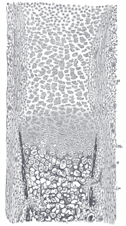
Section of fetal bone of cat. ir. Irruption of the subperiosteal tissue. p. Fibrous layer of the periosteum. o. Layer of osteoblasts. im. Subperiosteal bony deposit. Latin centrum ossificationis Gray's subject #18 93 The first step in ossification of the cartilage is that the cartilage cells, at the point where ossification is commencing and which is termed an ossification center, enlarge and arrange themselves in rows.[1]
The matrix in which they are imbedded increases in quantity, so that the cells become further separated from each other.
A deposit of calcareous material now takes place in this matrix, between the rows of cells, so that they become separated from each other by longitudinal columns of calcified matrix, presenting a granular and opaque appearance.
Here and there the matrix between two cells of the same row also becomes calcified, and transverse bars of calcified substance stretch across from one calcareous column to another.
Thus there are longitudinal groups of the cartilage cells enclosed in oblong cavities, the walls of which are formed of calcified matrix which cuts off all nutrition from the cells; the cells, in consequence, atrophy, leaving spaces called the primary areolæ.
Types of ossification centers
There are two types of ossification centers - primary and secondary.
A primary ossification center is the first area of a bone to start ossifying. It usually appears during prenatal development in the central part of each developing bone. In long bones the primary centers occur in the diaphysis/shaft and in irregular bones the primary centers occur usually in the body of the bone. Most bones have only one primary center (e.g. all long bones) but some irregular bones such as the os coxa (hip) and vertebrae have multiple primary centers.
A secondary ossification center is the area of ossification that appears after the primary ossification center has already appeared - most of which appear during the postnatal and adolescent years. Most bones have more than one secondary ossification center. In long bones, the secondary centres appear in the epiphyses.
References
- ^ Gray and Spitzka (1910), page 44.
Bibliography
- Gray, Henry; Spitzka, Edward Anthony (1910). Anatomy, descriptive and applied. the University of California: Lea & Febiger. http://books.google.com/books?id=a9wEAQAAIAAJ&pg=PA44&dq=ossification&hl=en&ei=e27HTe2AFoy5twe97tWfBA&sa=X&oi=book_result&ct=result&resnum=4&ved=0CDkQ6AEwAw#v=onepage&q=ossification&f=false.
This article was originally based on an entry from a public domain edition of Gray's Anatomy. As such, some of the information contained within it may be outdated.
Embryology of bones, joints, and muscles (GA 2.80, TE E5.0-2) Ossification Lower limbHeadcranium: Ossification of occipital bone · Ossification of frontal bone · Ossification of temporal bone · Ossification of sphenoid · Ossification of ethmoid
facial bones: Ossification of vomer (Sutura vomerina · Foramen vomerinum · Meatus vomerinus · Fissura vomerina) · Ossification of maxilla · Ossification of mandibleOtherOther M: JNT
anat(h/c, u, t, l)/phys
noco(arth/defr/back/soft)/cong, sysi/epon, injr
proc, drug(M01C, M4)
Categories:- Skeletal system
- Tissues
- Musculoskeletal system stubs
- Developmental biology stubs

Wikimedia Foundation. 2010.