- Erdheim–Chester disease
-
Erdheim-Chester disease Classification and external resources 
ICD-10 C96.1 ICD-9 202.3 DiseasesDB 29792 MeSH D031249 Erdheim–Chester disease (also known as Erdheim–Chester syndrome or polyostotic sclerosing histiocytosis) is a rare disease characterized by the abnormal multiplication of a specific type of white blood cells called histiocytes, or tissue macrophages (technically, this disease is termed a non-Langerhans-cell histiocytosis). Usually, onset is in middle age. The disease involves an infiltration of lipid-laden macrophages, multinucleated giant cells, an inflammatory infiltrate of lymphocytes and histiocytes in the bone marrow, and a generalized sclerosis of the long bones.[1]
Contents
History
The first case of ECD was reported by the American pathologist William Chester in 1930.[2]
Clinical presentation
ECD affects predominantly adults, with a mean age of 53 years.[3]
Long bone involvement is almost universal in ECD patients and is bilateral and symmetrical in nature. More than 50% of cases have some sort of extraskeletal involvement. This can include kidney, skin, brain and lung involvement, and less frequently retroorbital tissue, pituitary gland and heart involvement is observed. Bone pain is the most frequent of all symptoms associated with ECD and mainly affects the lower limbs, knees and ankles. The pain is often described as mild but permanent, and juxtaarticular in nature. Exophthalmos occurs in some patients and is usually bilateral, symmetric and painless. In most cases it occurs several years before the final diagnosis.
A review of 59 case studies by Veyssier-Belot, C et al. in 1996 reported the following symptoms in order of frequency of occurrence:[3]
- Bone pain
- Retroperitoneal fibrosis
- Diabetes insipidus
- Exophthalmos
- Xanthomas
- Neurological signs (including Ataxia)
- Dyspnea caused by interlobular septal and pleural thickening.
- Kidney Failure
- Hypopituitarism
- Liver Failure
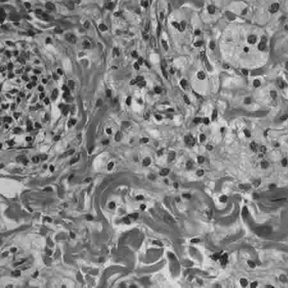
Histology
Histologically, ECD differs from Langerhans cell histiocytosis (LCH) in a number of ways. Unlike LCH, ECD does not stain positive for S-100 or CD 1a, and electron microscopy of cell cytoplasm does not disclose Birbeck granules.[3] Tissue samples show xanthomatous or xanthogranulomatous infiltration by lipid-laden or foamy histiocytes, and are usually surrounded by fibrosis. Bone biopsy is said to offer the greatest likelihood of reaching a diagnosis. So in a nutshell, there is histiocyte proliferation and on staining, the section is CD68+ and CD1a-
Diagnosis
Radiologic osteosclerosis and histology are the main diagnostic features. Diagnosis can often be difficult because of the rareness of ECD as well as the need to differentiate it from LCH. A diagnosis from neurological imaging may not be definitive. The presence of symmetrical cerebellar and pontine signal changes on T2-weighted images seem to be typical of ECD, however, multiple sclerosis and metabolic diseases must also be considered in the differential diagnosis.[4]
ECD is not a common cause of exophthalmos but can be diagnosed by biopsy. However, like all biopsies, this may be inconclusive.[5]
Treatment
Current treatment options include:
- Surgical Debulking
- High-dose Corticosteroid therapy
- Cyclosporine
- Interferon-α[5]
- Chemotherapy
- Radiation therapy
All current treatments have had varying degrees of success.
The vinca alkaloids and anthracyclines have been used most commonly in ECD treatment.[6]
Support groups
The Erdheim–Chester Disease Global Alliance is a support and advocacy group with the goal of raising awareness of and promoting research into ECD.[7]
References
- ^ "Erdheim–Chester disease at the United States National Library of Medicine". http://www.nlm.nih.gov/cgi/mesh/2003/MB_cgi?term=ERDHEIM-CHESTER+DISEASE. Retrieved 2008-06-19.
- ^ Chester W (1930). "Über Lipoidgranulomatose". Virchows Arch Pathol Anat Physiol. 279: 561–602.
- ^ a b c Veyssier-Belot C, Cacoub P, Caparros-Lefebvre D, et al. (1996). "Erdheim-Chester disease. Clinical and radiologic characteristics of 59 cases". Medicine (Baltimore) 75 (3): 157–69. PMID 8965684.
- ^ Weidauer S, von Stuckrad-Barre S, Dettmann E, Zanella FE, Lanfermann H (2003). "Cerebral Erdheim–Chester disease: case report and review of the literature". Neuroradiology 45 (4): 241–5. doi:10.1007/s00234-003-0950-z. PMID 12687308.
- ^ a b "Erdheim Chester Disease - M. D. Anderson Cancer Center". http://www.mdanderson.org/diseases/eyecancer/display.cfm?id=5423a04c-459e-4e12-a737309ef2e1e299&method=displayfull. Retrieved 2007-08-26.
- ^ Gupta A, Kelly B, McGuigan JE (2002). "Erdheim–Chester disease with prominent pericardial involvement: clinical, radiologic, and histologic findings". Am. J. Med. Sci. 324 (2): 96–100. doi:10.1097/00000441-200208000-00008. PMID 12186113. http://meta.wkhealth.com/pt/pt-core/template-journal/lwwgateway/media/landingpage.htm?issn=0002-9629&volume=324&issue=2&spage=96.
- ^ "Erdheim–Chester Disease". ECD Global Alliance. http://www.erdheim-chester.org. Retrieved 2009-05-08.
External links
- 01067 at CHORUS
- Writeup at Harvard University
Histiocytosis (D76.0, 277.89) WHO-I/Langerhans cell histiocytosis/
X-type histiocytosisLetterer–Siwe disease · Hand–Schüller–Christian disease · Eosinophilic granuloma · Congenital self-healing reticulohistiocytosisWHO-II/non-Langerhans cell histiocytosis/
Non-X histiocytosisJuvenile xanthogranuloma · Hemophagocytic lymphohistiocytosis · Erdheim-Chester disease · Niemann-Pick disease · Sea-blue histiocyte syndrome · Benign cephalic histiocytosis · Generalized eruptive histiocytoma · Xanthoma disseminatum · Progressive nodular histiocytosis · Papular xanthoma · Hereditary progressive mucinous histiocytosis · Reticulohistiocytosis (Multicentric reticulohistiocytosis, Reticulohistiocytoma) · Indeterminate cell histiocytosisWHO-III/malignant histiocytosis Histiocytic sarcoma · Langerhans cell sarcoma · Interdigitating dendritic cell sarcoma · Follicular dendritic cell sarcomaUngrouped Categories:- Rare diseases
- Histiocytosis
- Skeletal disorders
- Syndromes
Wikimedia Foundation. 2010.