- Dose-volume histogram
-
Dose-volume histogram (DVH), a concept used in radiation treatment planning[1]. DVHs were introduced by Goitein and Verhey in 1979 in a publication by Shipley et al.[2]. The purpose of a DVH is to summarize 3D dose distributions in a graphical 2D format. In modern radiation therapy, 3D dose distributions are typically created in a computerized treatment planning system based on a 3D reconstruction of a CT scan. The "volume" referred to in DVH analysis can be a target of radiation treatment, a healthy organ nearby a target, or an arbitrary structure.
DVHs can be visualized in either of two ways: differential DVHs or cumulative DVHs. A DVH is created by first determining the size of the dose bins of the histogram. Bins can be of arbitrary size, e.g. 0-1 Gy, 1.001-2 Gy, 2.001-3 Gy, etc. In a differential DVH, bar or column height indicates the volume of structure receiving a dose given by the bin. Bin doses are along the horizontal axis, and structure volumes (either percent or absolute volumes) are on the vertical. The differential DVH takes the appearance of a typical histogram. The cumulative DVH is plotted with bin doses along the horizontal axis, as well. However, the column height of the first bin (0-1 Gy, e.g.) represents the volume of structure receiving greater than or equal to that dose. The column height of the second bin (1.001-2 Gy, e.g.) represents the volume of structure receiving greater than or equal to that dose, etc. With very fine (small) bin sizes, the cumulative DVH takes on the appearance of a smooth line graph. The lines always slope and start from top-left to bottom-right. For a structure receiving a very homogenous dose—100% of the volume receiving exactly 10 Gy for example—the cumulative DVH will appear as a horizontal line at the top of the graph, at 100% volume as plotted vertically, with a vertical drop at 10 Gy on the horizontal axis.
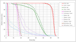 A cumulative DVH from a radiotherapy plan.
Cumulative DVHs are overwhelmingly used and preferred over differential DVHs. The DVH is ubiquitous in the medical specialty of radiation oncology. A DVH used clinically usually includes all structures and targets of interest in the radiotherapy plan, each line plotted a different color representing a different structure. The vertical axis is almost always plotted as percent volume (rather than absolute volume), as well.
Clinical studies have shown that DVH metrics correlate with patient toxicity outcomes[citation needed]. A drawback of the DVH methodology is that it offers no spatial information; i.e., a DVH does not show where within a structure a dose is received. Also, DVHs from initial radiotherapy plans represent the doses to structures at the start of radiation treatment. As treatment progresses and time elapses, if there are changes (i.e. if patients lose weight, if tumors shrink, if organs change shape, etc.), the original DVH loses validity.
References
Categories:- Radiation oncology
- Medical physics

Wikimedia Foundation. 2010.