- Frontalis muscle
Muscle infobox
Name = Frontalis
Latin = venter frontalis musculi occipitofrontalis
GraySubject = 105
GrayPage = 379
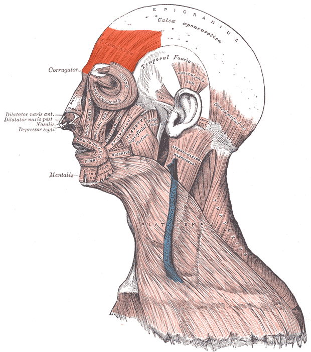
Caption = Visible at top left
Origin =galea aponeurosis
Insertion =Skin above the eyebrows
Blood =ophthalmic artery
Nerve =facial nerve
Action = wrinklesbrow
DorlandsPre = m_22
DorlandsSuf = 12549942
The Frontalismuscle is thin, of a quadrilateral form, and intimately adherent to thesuperficial fascia . It is broader than theOccipitalis and its fibers are longer and paler in color. It is located on the front of the head.It has no bony attachments.
Its medial fibers are continuous with those of the
Procerus ; its immediate fibers blend with the Corrugator andOrbicularis oculi , thus attached to the skin of the eyebrows [ "eye, human."Encyclopædia Britannica. 2008. Encyclopædia Britannica 2006 Ultimate Reference Suite DVD 5 Apr. 2008 ] ; and its lateral fibers are also blended with the latter muscle over thezygomatic process of thefrontal bone .In the eyebrows, its primary function is to lift them (thus opposing the orbital portion of the orbicularis), especially when looking up. It also acts when a view is too distant or dim. [ Ibid ]
From these attachments the fibers are directed upward, and join the
galea aponeurotica below thecoronal suture .The medial margins of the Frontales are joined together for some distance above the root of the
nose ; but between the Occipitales there is a considerable, though variable, interval, occupied by thegalea aponeurotica .It could be part of
occipitofrontalis muscle .
=AdditionalReferences
External links
*
*
* [http://www.ptcentral.com/muscles/musclehead.html#frontalis PTCentral]
Wikimedia Foundation. 2010.