- Brain size
-
Brain size is one aspect of animal anatomy and evolution. Both overall brain size and the size of substructures have been analysed, and the question of links between size and functioning - particularly intelligence - has often proved controversial. Brain size is sometimes measured by weight and sometimes by volume (via MRI scans or by skull volume).
Contents
Comparisons among animals
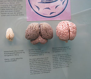
See also: Cetacean intelligence#Brain sizeScale model of bottlenose dolphin (Tursiops truncatus) brain (middle), compared with brains of wild pig (Sus scrofa) (left), and man (Homo sapiens) (right)
The largest brains are those of sperm whales, weighing about 8 kg (18 lbs). An elephant's brain weighs just over 5 kg (11 lbs), a bottlenose dolphin's 1.5 to 1.7 kg, whereas a human brain is around 1.3 to 1.5 kg (3 lbs).
Brain size tends to vary according to body size.
The relationship is not proportional, though: the brain-to-body mass ratio varies. The largest ratio found is in the shrew.[1] Averaging brain weight across all orders of mammals, it follows a power law, with an exponent of about 0.75.[2] There are good reasons to expect a power law: for example, the body-size to body-length relationship follows a power law with an exponent of 0.33, and the body-size to surface-area relationship a power law with an exponent of 0.67. The explanation for an exponent of 0.75 is not obvious; however, it is worth noting that several physiological variables appear to be related to body size by approximately the same exponent - for example, the basal metabolic rate.[3]
This power law formula applies to the "average" brain of mammals taken as a whole, but each family (cats, rodents, primates, etc.) departs from it to some degree, in a way that generally reflects the overall "sophistication" of behavior.[4] Primates, for a given body size, have brains 5 to 10 times as large as the formula predicts. Predators tend to have relatively larger brains than the animals they prey on; placental mammals (the great majority) have relatively larger brains than marsupials such as the opossum. A standard formula for assessing an animal's brain size compared to what would be expected from its body size, is known as the encephalization quotient. The encephalization quotient for humans is approximately 4.6.[5]
When the mammalian brain increases in size, not all parts increase at the same rate.[6] In particular, the larger the brain of a species, the greater the fraction taken up by the cortex. Thus, in the species with the largest brains, most of their volume is filled with cortex: this applies not only to humans, but also to animals such as dolphins, whales, or elephants.
The evolution of Homo sapiens over the past two million years has been marked by a steady increase in brain size, but much of it can be accounted for by corresponding increases in body size.[7] There are, however, many departures from the trend that are difficult to explain in a systematic way: in particular, the appearance of modern man about 100,000 years ago was marked by a decrease in body size at the same time as an increase in brain size. Even so, it is notorious that Neanderthals, which went extinct about 40,000 years ago, had larger brains than modern Homo sapiens.[8]
Not all investigators are happy with the amount of attention that has been paid to brain size. Roth and Dicke, for example, have argued that factors other than size are more highly correlated with intelligence, such as the number of cortical neurons and the speed of their connections.[9] Moreover they point out that intelligence depends not just on the amount of brain tissue, but on the details of how it is structured. It is also well known that crows, ravens, and African Grey Parrots are quite intelligent even though they have small brains.
While humans have the largest encephalization quotient of extant animals, it is not out of line for a primate. Gorillas are out of line, having a smaller brain to body ratio than would be expected.[10][11]
Some other anatomical trends are correlated in the human evolutionary path with brain size: the basicranium becomes more flexed with increasing brain size relative to basicranial length.[12]
Modern humans
The balance of findings, which have been largely on participants of European ancestry, indicate an average adult brain volume of 1130 cubic centimetres (cc) for women and 1260 cc for men. There is substantial variation however;[13] a study of 46 adults aged 22–49 years and of mainly European descent, found an average brain volume of 1273.6cc for men, ranging from 1052.9 to 1498.5cc, and 1131.1cc for women, ranging from 974.9 to 1398.1cc.[14] The right cerebral hemisphere is typically larger than the left, whereas the cerebellar hemispheres are typically of more similar size.
Evolution: the paradox of Homo floresiensis
 Evolution of brain volume in homininaee
There has been a gradual increase in brain volume as we progressed along the Human timeline of evolution (see Homininae), starting from about 600 cm3 in Homo habilis up to 1500 cm3 in Homo sapiens neanderthalensis. Thus, in general there's a correlation between brain volume and intelligence. However, modern Homo sapiens have a brain volume slightly smaller (1250 cm3) than neanderthals, women have a brain volume slightly smaller than men (see before) and the Flores island hominins (Homo floresiensis), nicknamed hobbits, had a cranial capacity of about 380 cm3 (considered small for a chimpanzee) about a third of that of H. erectus. It is proposed that they evolved from H. erectus as a case of insular dwarfism. With their three times smaller brain the Flores hominids apparently used fire and made stone tools at least as sophisticated as those of their ancestor H.erectus. In this case, it seems that for intelligence, the structure of the brain is more important than its size.
Age and Sex
Overall, there is a background of similarity between adult brain volume measures of people of differing ages and sexes. Nevertheless, underlying structural asymmetries do exist. Males have been found to have on average greater cerebral, cerebellar and cerebral cortical lobar volumes, except possibly left parietal.[15] The gender differences in size vary by more specific brain regions. Studies have tended to indicate that men have a relatively larger amygdalae and hypothalamus, while women have a relatively larger caudate and hippocampi. When covaried for intracranial volume, height, and weight, the balance of studies indicates women have a higher percentage of gray matter, whereas men have a higher percentage of white matter and cerebrospinal fluid. There is high variability between individuals in these studies, however.[13]
Genetic contribution
Adult twin studies have indicated high heritability estimates for overall brain size in adulthood (between 66% and 97%). The effect varies regionally within the brain, however, with high heritabilities of frontal lobe volumes (90-95%), moderate estimates in the hippocampi (40-69%), and environmental factors influencing several medial brain areas. In addition, lateral ventricle volume appears to be mainly explained by environmental factors, suggesting such factors also play a role in the surrounding brain tissue. Genes may cause the association between brain structure and cognitive functions, or the latter may influence the former during life. A number of candidate genes have been identified or suggested, but await replication.[16][17]
Neuroplasticity
A discovery in recent years is that the structure of the adult human brain changes when a new cognitive or motor skill, including vocabulary, is learned.[18] Structural neuroplasticity (increased grey matter volume) has been demonstrated in adults after three months of training in a visual-motor skill, with the qualitative change (i.e. learning of a new task) appearing more critical for the brain to change its structure than continued training of an already-learned task. Such changes (e.g. revising for medical exams) have been shown to last for at least 3 months without further practicing; other examples include learning novel speech sounds, musical ability, navigation skills and learning to read mirror-reflected words.[19][20]
Intelligence
See also: Neuroscience and intelligence#Brain size
Studies have tended to indicate small to moderate correlations (averaging around 0.3 to 0.4) between brain volume and IQ. The most consistent associations are observed within the frontal, temporal, and parietal lobes, the hippocampi, and the cerebellum, but only account for a relatively small amount of variance in IQ, which itself only shows a partial relationship to the general concept of intelligence and real-world performance.[21][22] In addition, brain volumes do not correlate strongly with other and more specific cognitive measures.[14] In men, IQ correlates more with gray matter volume in the frontal lobe and parietal lobe, whereas in women it correlates with gray matter volume in the frontal lobe and Broca's area, which is involved in language.[13]
Development and Aging
There is variation in child development in the size of different brain structures between individuals and genders.[23] Total cerebral and grey matter volumes peak during the ages from 10–20 years (earlier in girls than boys), whereas white matter and ventricular volumes increase. There is a general pattern in neural development of childhood peaks followed by adolescent declines (e.g. synaptic pruning). Consistent with adult findings, average cerebral volume is approximately 10% larger in boys than girls. However, such differences should not be interpreted as imparting any sort of functional advantage or disadvantage; gross structural measures may not reflect functionally relevant factors such as neuronal connectivity and receptor density, and of note is the high variability of brain size even in narrowly defined groups, for example children at the same age may have as much as a 50% differences in total brain volume.[24] Young girls have on average relative larger hippocampal volume, whereas the amygdalae are larger in boys.[13]
Significant dynamic changes in brain structure take place through adulthood and aging, with substantial variation between individuals. In later decades, men show greater volume loss in whole brain volume and in the frontal lobes, and temporal lobes, whereas in women there is increased volume loss in the hippocampi and parietal lobes.[13] Men show a steeper decline in global grey matter volume, although in both sexes it varies by region with some areas exhibiting little or no age effect. Overall white matter volume does not appear to decline with age, although there is variation between brain regions.[25]
See also
- Cranial capacity (should perhaps be merged with this article)
- Craniometry — includes historical discussion
- Neuroscience and intelligence
Notes
- ^ Brains of White Matter
- ^ Armstrong, 1983
- ^ Savage et al., 2004
- ^ Jerison, Evolution of the Brain and Intelligence
- ^ Aiello & Wheeler, 1995
- ^ Finlay et al., 2001
- ^ Kappelman, 1993
- ^ Holloway, 1995
- ^ Roth & Dicke, 2005
- ^ "Size isn't everything: The big brain myth" by Alison Motluk, New Scientist, July 31, 2010, pp. 38-41.
- ^ "Equal numbers of neuronal and nonneuronal cells make the human brain an isometrically scaled-up primate brain", by Frederico Azevedo et al., The Journal of Comparative Neurology, vol 513, p 532.
- ^ Ross & Henneberg, 1995
- ^ a b c d e Cosgrove et al., 2007
- ^ a b Allen et al., 2002
- ^ Carne et al., 2006
- ^ Peper, 2007
- ^ Zhang, 2003
- ^ Lee et al., 2007
- ^ Driemeyer et al., 2008
- ^ Ilg et al., 2008
- ^ Luders et al., 2008
- ^ Hoppe & Stojanovic, 2008
- ^ Lange et al., 1997
- ^ Giedd, 2008
- ^ Good et al., 2001
References
- Aiello, L; Wheeler, P (1995). "The Expensive Tissue Hypothesis: The Brain and the Digestive System in Human and Primate Evolution". Current Anthropology 36 (2): 199–221. doi:10.1086/204350. http://people.biology.ufl.edu/sphelps/documents/evobrain/aeillo_wheeler_95.pdf. Retrieved 15 April 2011.
- Allen, JS; Damasio H, Grabowski TJ (2002). "Normal neuroanatomical variation in the human brain: An MRI-volumetric study". Am J Phys Anthropol 118 (4): 341–58. doi:10.1002/ajpa.10092. PMID 12124914. http://www3.interscience.wiley.com/journal/96515947/abstract.
- Armstrong, E (1983). "Relative brain size and metabolism in mammals". Science 220 (4603): 1302–4. doi:10.1126/science.6407108. PMID 6407108.
- Carne, RP; Vogrin S, Litewka L, Cook MJ (2006). "Cerebral cortex: An MRI-based study of volume and variance with age and sex". J Clin Neurosci 13 (1): 60–72. doi:10.1016/j.jocn.2005.02.013. PMID 16410199.
- Cosgrove, KP; Mazure CM, Staley JK (2007). "Evolving Knowledge of Sex Differences in Brain Structure, Function and Chemistry". Biol Psychiat 62 (8): 847–55. doi:10.1016/j.biopsych.2007.03.001. PMC 2711771. PMID 17544382. http://linkinghub.elsevier.com/retrieve/pii/S0006322307001989.
- Driemeyer, J; Boyke, J, Gaser, C, Buchel, C, May, A (2008). Eagleman, David M.. ed. "Changes in Gray Matter Induced by Learning—Revisited". PLoS ONE 3 (7): 7. doi:10.1371/journal.pone.0002669. PMC 2447176. PMID 18648501. http://www.pubmedcentral.nih.gov/articlerender.fcgi?tool=pmcentrez&artid=2447176.
- Finlay, BL; Darlington RB, Nicastro N (2001). "Developmental structure in brain evolution" (PDF). Behav Brain Sci 20 (2): 263–308. PMID 11530543. http://anthropology.emory.edu/FACULTY/ANTJR/pdf/BBS.pdf.
- Giedd, JN (2008). "The teen brain: insights from neuroimaging". J Adolescent Health 42 (4): 335–43. doi:10.1016/j.jadohealth.2008.01.007. PMID 18346658. http://anthropology.emory.edu/FACULTY/ANTJR/pdf/BBS.pdf.
- Good, CD; Johnsrude IS, Ashburner J, Henson RN, Friston KJ, Frackowiak RS (2001). "A voxel-based morphometric study of ageing in 465 normal adult human brains". NeuroImage 14 (1 Pt 1): 21–36. doi:10.1006/nimg.2001.0786. PMID 11525331.
- Holloway, RL (1995). Changeaux JP, Chavillon J. ed. Origins of the Human Brain. Clarendon. pp. 42–54. ISBN 9780198523079. http://books.google.com/?id=SDmjJwAACAAJ.
- Ilg, R; Wohlschläger AM, Gaser C, Liebau Y, Dauner R, Wöller A, Zimmer C, Zihl J, Mühlau M (2008). "Gray matter increase induced by practice correlates with task-specific activation: a combined functional and morphometric magnetic resonance imaging study". J Neurosci 28 (16): 4210–5. doi:10.1523/JNEUROSCI.5722-07.2008. PMID 18417700.
- Jerison, HJ (1973). Evolution of the Brain and Intelligence. Academic Press. ISBN 9780123852502. http://books.google.com/?id=xTpDAAAACAAJ.
- Kappelman, J (1993). "The evolution of body mass and relative brain size in fossil hominids". J Human Evol 30 (3): 243–76. doi:10.1006/jhev.1996.0021.
- Lange, N; Giedd JN, Castellanos FX, Vaituzis AC, Rapoport JL (1997). "Variability of human brain structure size: ages 4–20 years". Psychiat Res: Neuroimaging 74 (6): 1–12. doi:10.1016/S0925-4927(96)03054-5. PMID 10710158.
- Lee, H; Devlin JT, Shakeshaft C, Stewart LH, Brennan A, Glensman J, Pitcher K, Crinion J, Mechelli A, Frackowiak RS, Green DW, Price CJ (2007). "Anatomical traces of vocabulary acquisition in the adolescent brain". J Neurosci 27 (5): 1184–9. doi:10.1523/JNEUROSCI.4442-06.2007. PMID 17267574. http://www.jneurosci.org/cgi/content/abstract/27/5/1184.
- Hoppe, C; Stojanovic J (2008). "High-aptitude minds: the neurological roots of genius". Scientific American. http://www.sciam.com/article.cfm?id=high-aptitude-minds.
- Luders, E; Narr KL, Thompson PM, Toga AW (2008). "Neuroanatomical Correlates of Intelligence". Intelligence 37 (2): 156–163. doi:10.1016/j.intell.2008.07.002. PMC 2770698. PMID 20160919. http://www.pubmedcentral.nih.gov/articlerender.fcgi?tool=pmcentrez&artid=2770698.
- Peper, JS; Brouwer, RM; Boomsma, DI; Kahn, RS; Hulshoff Pol, HE (2007). "Genetic influences on human brain structure: A review of brain imaging studies in twins". Human Brain Mapping 28 (6): 464–73. doi:10.1002/hbm.20398. PMID 17415783.
- Ross, C; Henneberg M (1995). "Basicranial flexion, relative brain size, and facial kyphosis in Homo sapiens and some fossil hominids". Am J Phys Anthropol 98 (4): 575–93. doi:10.1002/ajpa.1330980413. PMID 8599387. http://rosslab.uchicago.edu/pdf/rossandhenneberg1995.pdf.
- Roth, G; Dicke U (2005). "Evolution of the brain and intelligence". Trends Cogn Sci 9 (5): 250–7. doi:10.1016/j.tics.2005.03.005. PMID 15866152. http://www.subjectpool.com/ed_teach/y3project/Roth2005_TICS_brain_size_and_intelligence.pdf.
- Savage, MV; Gillooly JF, Woodruff WH, West GB, Allen AP, Enquist BJ, Brown JH (2004). "The predominance of quarter-power scaling in biology". Functional Ecol 18 (2): 257–82. doi:10.1111/j.0269-8463.2004.00856.x.
- Zhang, J (2003). "Evolution of the human ASPM gene, a major determinant of brain size". Genetics 165 (4): 2063–70. PMC 1462882. PMID 14704186. http://www.pubmedcentral.nih.gov/articlerender.fcgi?tool=pmcentrez&artid=1462882.
Categories:
Wikimedia Foundation. 2010.

