- Canthus (anatomy)
Infobox Anatomy
Name = PAGENAME
Latin =
GraySubject =
GrayPage =
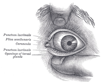
Caption = Front of lefteye witheyelids separated to show medialcanthus .
Caption2 =
Precursor =
System =
Artery =
Vein =
Nerve =
Lymph =
MeshName =
MeshNumber =
DorlandsPre = c_05
DorlandsSuf = 12210029Canthus (pl. canthi, palpebral commissures) is either corner of the
eye where the upper and lowereyelid s meet.The bicanthal plane is the
transversal plane linking both canthi and defines the upper boundary of themidface .Commissures
* The "lateral palpebral commissure" (commissura palpebrarum lateralis; external canthus) is more acute than the medial, and the eyelids here lie in close contact with the bulb of the eye.
* The "medial palpebral commissure" (commissura palpebrarum medialis; internal canthus) is prolonged for a short distance toward the
nose , and the two eyelids are separated by a triangular space, the lacus lacrimalis.urgery
Canthoplasty refers to a plastic surgery of the medial and/or lateral canthus.
A
canthotomy involves cutting the canthus, often performed to release excessive orbital pressure (i.e., from orbital hemorrhage or infection).Pathology
"Dystopia canthorum" is associated with
Waardenburg syndrome .ee also
*
Epicanthal fold External links
* [http://www.sheinman.com/Aanatomyp1.htm Diagram at sheinman.com]
* [http://www.solobambini.com/measure_canthus.html Diagram at solobambini.com] (measure ofPupillary distance )
Wikimedia Foundation. 2010.