- Trapezoid body
Infobox Brain
Name = PAGENAME
Latin = corpus trapezoideum
GraySubject = 191
GrayPage = 858
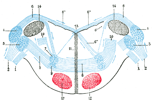
Caption = Terminal nuclei of thecochlear nerve , with their upper connections. (Schematic.) The vestibular nerve with its terminal nuclei and their efferent fibers have been suppressed. On the other hand, in order not to obscure the trapezoid body, the efferent fibers of the terminal nuclei on the right side have been resected in a considerable portion of their extent. The trapezoid body, therefore, shows only one-half of its fibers, viz., those which come from the left. 1.Vestibular nerve , divided at its entrance into themedulla oblongata . 2.Cochlear nerve . 3.Accessory nucleus of acoustic nerve. 4.Tuberculum acusticum . 5. Efferent fibers of accessory nucleus. 6. Efferent fibers oftuberculum acusticum , forming thestriae medullares , with 6’, their direct bundle going to thesuperior olivary nucleus of the same side; 6’’, their decussating bundles going to the superior olivary nucleus of the opposite side. 7. Superior olivary nucleus. 8.Trapezoid body . 9.Trapezoid nucleus . 10.Central acoustic tract (lateral lemniscus ). 11.Raphé . 12.Cerebrospinal fasciculus . 13.Fourth ventricle . 14.Inferior peduncle .
Caption2 =
IsPartOf =
Components =
Artery =
Vein =
Acronym =
BrainInfoType = hier
BrainInfoNumber = 589
MeshName =
MeshNumber =
DorlandsPre = c_56
DorlandsSuf = 12260860
The trapezoid body is part of theacoustic pathway . It is a bundle of fibers and cells in thepontine tegmentum . It consists of fibers arising from the ventralcochlear nucleus . A collection of nerve cells inside forms atrapezoid nucleus . Thesuperior olivary nucleus is situated on the dorsal surface of the trapezoid body. Most nerve fibers pass directly from the superior olivary nuclei to theinferior colliculus .Axons leaving the
ventral cochlear nucleus (VCN) form a broad pathway that crosses under the brain stem in the trapezoid body. A thin pathway, the intermediate acoustic stria, also leaves the VCN, merging with the trapezoid body close to thesuperior olivary complex , where many of its axons synapse. Axons leaving thedorsal cochlear nucleus (DCN) form the dorsal acoustic stria, which reaches primarily the contralateral dorsal nucleus of thelateral lemniscus and the central nucleus of the inferior colliculus.
Wikimedia Foundation. 2010.