- Sestamibi
drugbox
IUPAC_name = -

imagename = 99mTc-sestamibi
CAS_number = 109581-73-9
ATC_prefix = V09
ATC_suffix = GA01
PubChem = 5384
DrugBank =
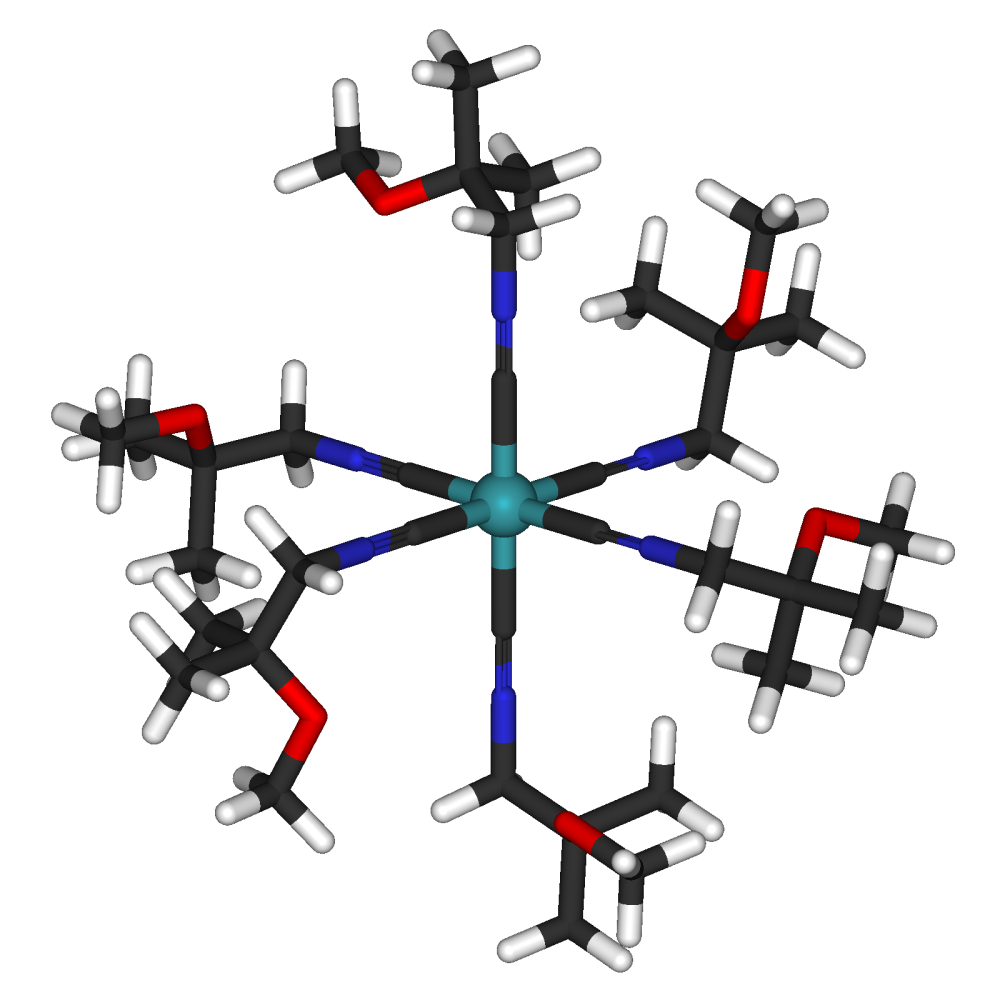
C = 36 | H = 66 | N = 6 | O = 6 | Tc = 1
molecular_weight = 777.852 g/mol
bioavailability = NA
protein_bound = 1%
metabolism = Nil
elimination_half-life = Variable
excretion = Fecal (33%) and renal (27%)
pregnancy_AU =
pregnancy_US = C
pregnancy_category =
licence_US = TECHNETIUM_TC-99M_SESTAMIBI_KIT
legal_AU =
legal_CA =
legal_UK =
legal_US = Rx-only
legal_status =
routes_of_administration = IntravenousSestamibi is a
radiopharmaceutical used innuclear medicine imaging. It is also known as methoxyisobutylisonitrile or MIBI and is sold under the brand name Cardiolite. The generic became available late September of 2008. Theradioisotope attached to the sestamibi molecule istechnetium -99m, forming 99mTc-sestamibi (or Tc99m MIBI).Its main use is for imaging the
myocardium (heart muscle). It is also used in the work-up of primaryhyperparathyroidism to identifyparathyroid adenomas, forradioguided surgery of the parathyroid and in the work-up of possible breast malignancies.Cardiac imaging
Technetium-99m sestamibi is a
lipophilic cation which, when injected intravenously into a patient, distributes in the myocardium proportionally to the myocardial blood flow. As opposed toThallium -201, MIBI does not undergo significant redistribution. Single photon emission computed tomography (SPECT ) imaging of the heart is performed using agamma camera to detect thegamma ray s emitted by the technetium-99m sestamibi as it decays. Two sets of images are acquired. For one set, the Tc99m MIBI is injected while the patient is at rest and then the myocardium is imaged. In the second set, the patient is stressed either by exercising on a treadmill or pharmacologically. The Tc99m MIBI is injected at peak stress and then imaging is performed . The resulting two sets of images are compared with each other to distinguish ischemic from infarcted areas of the myocardium. This imaging technique is also known as myocardial perfusion imaging (MPI).Parathyroid imaging
In primary hyperparathyroidism, one or more of the four parathyroid glands either develops a benign tumor called an
adenoma or undergoes hypertrophy as a result of homeostatic dysregulation. The parathyroid gland take up Tc99m MIBI following an intravenous injection, and the patient's neck is imaged with a gamma camera to show the location of all glands. A second image is obtained after a washout time (approximately 2 hours), and mitochondria in the oxyphil cells of the abnormal glands retaining the Tc99m are seen with the gamma camera. This imaging method will detect 75 to 90 percent of abnormal parathyroid glands in primary hyperparathyroidism. Anotolaryngologist can then perform a directed parathyroidectomy (less invasive than traditional surgery) to remove the abnormal gland.Breast imaging
Tc99m MIBI is also used in the evaluation of breast nodules. Malignant breast tissues concentrate MIBI to a much greater extent and more frequently than benign disease. As such, limited characterization of breast anomalies is possible. Scintimammography
Breast-Specific Gamma Imaging (BSGI) has the highest specificity for breast cancer of any imaging test, and has a sensitivity of 66% based on positive biopsy compared to mammography and ultrasound with a 29% positive biopsy.Fact|date=January 2008Radioguided surgery of the parathyroids
Following the administration of Tc99m MIBI it collects in overactive parathyroid glands. During
surgery , the surgeon can use a probe sensitive to gamma rays to locate the overactive parathyroid before removing it. [ [http://www.springerlink.com/content/28up57k554526l67/ SpringerLink - Journal Article ] ]References
* [http://www.cardiolite.com/index_flash.html Cardiolite.com]
* [http://www.parathyroid.com/index.htm Parathyroid.com]
* [http://www.amershamhealth-us.com/myoview/ Myoview.com]
* [http://www.breastcancer.org/symptoms/testing/sestamibi.jsp Miraluma]
* [http://www.dilon.com/bsgi.php Dilon Breast-Specific Gama Imaging]

Wikimedia Foundation. 2010.