- Mitral valve prolapse
-
Mitral valve prolapses Classification and external resources 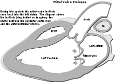
In mitral valve prolapse, the leaflets of the mitral valve prolapse back into the left atrium.ICD-10 I34.1 ICD-9 394.0, 424.0 OMIM 157700 DiseasesDB 8303 MedlinePlus 000180 eMedicine emerg/316 MeSH D008945 Mitral valve prolapse (MVP) is a valvular heart disease characterized by the displacement of an abnormally thickened mitral valve leaflet into the left atrium during systole.[1] There are various types of MVP, broadly classified as classic and nonclassic. In its nonclassic form, MVP carries a low risk of complications. In severe cases of classic MVP, complications include mitral regurgitation, infective endocarditis, congestive heart failure, and—in rare circumstances—cardiac arrest, usually resulting in sudden death.
The diagnosis of MVP depends upon echocardiography, which uses ultrasound to visualize the mitral valve. Early studies estimated a prevalence of 38% among healthy teenagers; with improved echocardiographic techniques and clear diagnostic criteria, the true prevalence of MVP is estimated at 2-3% of the population.[1]
The condition was first described by John Brereton Barlow in 1966. As such, it mayalso be referred to as Barlow's Syndrome.[2] and was subsequently termed mitral valve prolapse by J. Michael Criley.[3]
Contents
Overview
 St. Zenon of Verona wearing a mitre.
The mitral valve, so named because of its resemblance to a bishop's mitre, is the heart valve that prevents the backflow of blood from the left ventricle into the left atrium of the heart. It is composed of two leaflets, one anterior and one posterior, that close when the left ventricle contracts.
Each leaflet is composed of three layers of tissue: the atrialis, fibrosa, and spongiosa. Patients with classic mitral valve prolapse have excess connective tissue that thickens the spongiosa and separates collagen bundles in the fibrosa. This is due to an excess of dermatan sulfate, a glycosaminoglycan. This weakens the leaflets and adjacent tissue, resulting in increased leaflet area and elongation of the chordae tendineae. Elongation of the chordae tendineae often causes rupture, commonly to the chordae attached to the posterior leaflet. Advanced lesions—also commonly involving the posterior leaflet—lead to leaflet folding, inversion, and displacement toward the left atrium.[4]
Subtypes
 Diagnosis of mitral valve prolapse is based on modern echocardiographic techniques which can pinpoint abnormal leaflet thickening and other related pathology.
Prolapsed mitral valves are classified into several subtypes, based on leaflet thickness, concavity, and type of connection to the mitral annulus. Subtypes can be described as classic, nonclassic, symmetric, asymmetric, flail, or non-flail.[4]
All measurements below refer to adult patients; applying them to children may be misleading.
Classic versus nonclassic
Prolapse occurs when the mitral valve leaflets are displaced more than 2 mm above the mitral annulus high points. The condition can be further divided into classic and nonclassic subtypes based on the thickness of the mitral valve leaflets: up to 5 mm is considered nonclassic, while anything beyond 5 mm is considered classic MVP.[4]
Symmetric versus asymmetric
Classical prolapse may be subdivided into symmetric and asymmetric, referring to the point at which leaflet tips join the mitral annulus. In symmetric coaptation, leaflet tips meet at a common point on the annulus. Asymmetric coaptation is marked by one leaflet displaced toward the atrium with respect to the other. Patients with asymmetric prolapse are susceptible to severe deterioration of the mitral valve, with the possible rupture of the chordae tendineae and the development of a flail leaflet.[4]
Flail versus non-flail
Asymmetric prolapse is further subdivided into flail and non-flail. Flail prolapse occurs when a leaflet tip turns outward, becoming concave toward the left atrium, causing the deterioration of the mitral valve. The severity of flail leaflet varies, ranging from tip eversion to chordal rupture. Dissociation of leaflet and chordae tendineae provides for unrestricted motion of the leaflet (hence "flail leaflet"). Thus patients with flail leaflets have a higher prevalence of mitral regurgitation than those with the non-flail subtype.[4]
Diagnosis
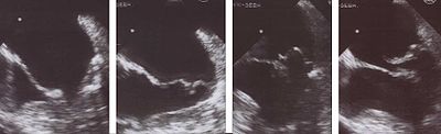 Transesophageal echocardiogram of mitral valve prolapse.
Echocardiography is the most useful method of diagnosing a prolapsed mitral valve. Two- and three-dimensional echocardiography are particularly valuable as they allow visualization of the mitral leaflets relative to the mitral annulus. This allows measurement of the leaflet thickness and their displacement relative to the annulus. Thickening of the mitral leaflets >5 mm and leaflet displacement >2 mm indicates classic mitral valve prolapse.[4]
Epidemiology
Prior to the strict criteria for the diagnosis of mitral valve prolapse, as described above, the incidence of mitral valve prolapse in the general population varied greatly.[4] Some studies estimated the incidence of mitral valve prolapse at 5 to 15 percent or even higher.[5] One study suggested MVP in up to 38% of healthy teenagers.[6]
Recent elucidation of mitral valve anatomy and the development of three-dimensional echocardiography have resulted in improved diagnostic criteria, and the true prevalence of MVP based on these criteria is estimated at 2-3%.[1] As part of the Framingham Heart Study, for example, the prevalence of mitral valve prolapse in Framingham, MA was estimated at 2.4%. There was a near-even split between classic and nonclassic MVP, with no significant age or sex discrimination.[7] MVP is observed in 7% of autopsies in the United States.[8]
Risk factors
MVP may occur with greater frequency in individuals with Ehlers-Danlos Syndrome, Marfan syndrome [9] or polycystic kidney disease.[10] Other risk factors include Graves' disease.[11] and chest wall deformities such as Pectus Excavatum [12]
Signs and symptoms
A correlation has been reported between bipolar disorder and mitral valve prolapse.[13]
For unknown reasons, MVP patients tend to have a low body mass index (BMI) and are typically leaner than individuals without MVP.[4][7]
Murmur
Upon auscultation of an individual with mitral valve prolapse, a mid-systolic click, followed by a late systolic murmur heard best at the apex is common.
In contrast to most other heart murmurs, the murmur of mitral valve prolapse is accentuated by standing and valsalva maneuver (earlier systolic click and longer murmur) and diminished with squatting (later systolic click and shorter murmur). The only other heart murmur that follows this pattern is the murmur of hypertrophic cardiomyopathy. A MVP murmur can be distinguished from a hypertrophic cardiomyopathy murmur by 1) the presence of a mid-systolic click which is virtually diagnostic of MVP, and 2) the fact that hand grip maneuver intensifies the murmur of MVP[citation needed] and diminishes the murmur of hypertrophic cardiomyopathy. The hand grip maneuver also diminishes the duration of the murmur and delays the timing of the mid-systolic click.[14]
Both valsalva maneuver and standing decrease venous return to the heart thereby decreasing left ventricular diastolic filling (preload) and causing more laxity on the chordae tendineae. This allows the mitral valve to prolapse earlier in systole, leading to an earlier systolic click (i.e. closer to S1), and a longer murmur. Hand grip maneuver increases total peripheral resistance (afterload) and therefore increases back pressure on the mitral valve resulting in a more intense murmur without changing the timing of the systolic click.
Mitral valve prolapse syndrome
Historically, the term mitral valve prolapse syndrome has been applied to MVP associated with palpitations, atypical chest pain, dyspnea on exertion, low body mass index, and electrocardiogram abnormalities in the setting of anxiety, syncope, low blood pressure, and other signs suggestive of autonomic nervous system dysfunction.[1] Modern studies, however, have failed to demonstrate that these features occur with greater frequency in individuals with MVP.[1]
Occasionally, supraventricular arrhythmias observed in MVP are associated with increased parasympathetic tone.[15]
Complications
Mitral regurgitation
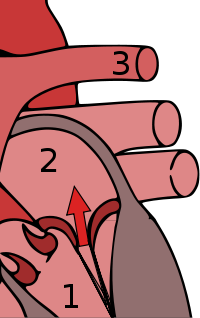 Mitral valve prolapse can result in mitral regurgitation, shown here, in which blood abnormally flows from the left ventricle into the left atrium.
Mitral valve prolapse is frequently associated with mild mitral regurgitation,[16] where blood aberrantly flows from the left ventricle into the left atrium during systole. In the United States, MVP is the most common cause of severe, non-ischemic mitral regurgitation.[1] This is occasionally due to rupture of the chordae tendineae that support the mitral valve. [14]
Sudden death
The MVP complications mitral regurgitation and congestive heart failure may, in turn, cause arrhythmias and atrial fibrillation that may progress and lead to sudden death. However, there is no evidence that a prolapsed valve itself contributes to such arrythmias.[17]
Prognosis
Generally, MVP is a benign disorder. However, MVP patients with a murmur, not just an isolated click, have a general mortality rate that is increased by 15-20%.[8] The major predictors of mortality are the severity of mitral regurgitation and the ejection fraction.[18]
Treatment
Most patients benefit from a nutritious diet, regular exercise, and a supportive environment.[19] Those with mitral valve prolapse and symptoms of dysautonomia (palpitations, chest pain) may often benefit from beta-blockers (e.g., propranolol). Patients with prior stroke and/or atrial fibrillation may require blood thinners, such as aspirin or warfarin. However, although beta blockers are commonly prescribed, they may be of limited benefit.[20] Unless otherwise contraindicated, many people with MVPS benefit from non-drug interventions, such as increased fluid and sodium intake, avoiding adrenalin-like substances found in many over-the-counter medications, avoiding caffeine, and getting regular cardiovascular exercise.[21]
Mitral valve prolapse associated with severe mitral regurgitation can be treated with repair or surgical replacement of the mitral valve. Repair of the mitral valve is always preferable to replacement and should be performed by surgeons that are skilled in the procedure. Current ACC/AHA guidelines suggest that early repair of mitral valve, performed in centers of surgical excellence, should be considered even in patients without symptoms of heart failure. Symptomatic patients, those with evidence of diminished left ventricular function or left ventricular dilatation need urgent attention.
Treatment with magnesium supplements may help reduce symptoms of MVP.[22]
Prevention of infective endocarditis
Individuals with MVP are at higher risk of bacterial infection of the heart, called infective endocarditis. This risk is approximately three- to eightfold the risk of infective endocarditis in the general population.[1] Until 2007, the American Heart Association recommended prescribing antibiotics before invasive procedures, including those in dental surgery. Thereafter, they concluded that "prophylaxis for dental procedures should be recommended only for patients with underlying cardiac conditions associated with the highest risk of adverse outcome from infective endocarditis." [23]
History
The term mitral valve prolapse was coined by J. Michael Criley in 1966 and gained acceptance over the other descriptor of "billowing" of the mitral valve, as described by John Brereton Barlow.[24]
References
- ^ a b c d e f g Hayek E, Gring CN, Griffin BP (2005). "Mitral valve prolapse". Lancet 365 (9458): 507–18. doi:10.1016/S0140-6736(05)17869-6. PMID 15705461.
- ^ Barlow JB, Bosman CK (February 1966). "Aneurysmal protrusion of the posterior leaflet of the mitral valve. An auscultatory-electrocardiographic syndrome". Am. Heart J. 71 (2): 166–78. doi:10.1016/0002-8703(66)90179-7. PMID 4159172.
- ^ Criley JM, Lewis KB, Humphries JO, Ross RS (July 1966). "Prolapse of the mitral valve: clinical and cine-angiocardiographic findings". Br Heart J 28 (4): 488–96. doi:10.1136/hrt.28.4.488. PMC 459076. PMID 5942469. http://www.pubmedcentral.nih.gov/articlerender.fcgi?tool=pmcentrez&artid=459076.
- ^ a b c d e f g h Playford, David; Weyman, Arthur (2001). "Mitral valve prolapse: time for a fresh look". Reviews in Cardiovascular Medicine 2 (2): 73–81. PMID 12439384. http://www.medreviews.com/pubmed.cfm?j=2&v=2&i=2&p=73.
- ^ Levy D, Savage D. (1987). "Prevalence and clinical features of mitral valve prolapse". Am Heart J 113 (5): 1281–90. doi:10.1016/0002-8703(87)90956-2. PMID 3554946.
- ^ Warth DC, King ME, Cohen JM, Tesoriero VL, Marcus E, Weyman AE (May 1985). "Prevalence of mitral valve prolapse in normal children". Journal of the American College of Cardiology 5 (5): 1173–7. doi:10.1016/S0735-1097(85)80021-8. PMID 3989128.
- ^ a b Freed LA, Levy D, Levine RA, Larson MG, Evans JC, Fuller DL, Lehman B, Benjamin EJ. (1999). "Prevalence and clinical outcome of mitral-valve prolapse". N Engl J Med 341 (1): 1–7. doi:10.1056/NEJM199907013410101. PMID 10387935.
- ^ a b Mitral Valve Prolapse at eMedicine
- ^ "Related Disorders: Mitral Valve Prolapse". Archived from the original on 2007-02-25. http://web.archive.org/web/20070225111027/http://www.marfan.org/nmf/GetContentRequestHandler.do?menu_item_id=80. Retrieved 2007-07-11.
- ^ Lumiaho, A; Ikäheimo, R; Miettinen, R; Niemitukia, L; Laitinen, T; Rantala, A; Lampainen, E; Laakso, M et al. (2001). "Mitral valve prolapse and mitral regurgitation are common in patients with polycystic kidney disease type 1.". American journal of kidney diseases 38 (6): 1208–16. doi:10.1053/ajkd.2001.29216. PMID 11728952.
- ^ "Mount Sinai - Disease". http://www.mountsinai.org/Patient%20Care/Service%20Areas/Heart/Diseases%20and%20Conditions?citype=Disease&ciid=Barlow%27s%20syndrome.
- ^ "Pectus Excavatum". http://emedicine.medscape.com/article/1004953-overview.
- ^ Giannini AJ, Price WA, Loiselle RH. (Aug 1984). "Prevalence of mitral valve prolapse in bipolar affective disorder". Am J Psychiatry 141 (8): 991–2. PMID 6465378.
- ^ a b Tanser, Paul H. (reviewed Mar 2007). "Mitral Valve Prolapse", The Merck Manuals Online Medical Library, Retrieved 2011-01-08.
- ^ Terechtchenko L, Doronina SA, Pochinok EM, Riftine A. (2003). "Autonomic tone in patients with supraventricular arrhythmia associated with mitral valve prolapse in young men". Pacing Clin Electrophysiol 26 (1 Pt 2): 444–6. doi:10.1046/j.1460-9592.2003.00067.x. PMID 12687863.
- ^ Kolibash AJ (1988). "Progression of mitral regurgitation in patients with mitral valve prolapse". Herz 13 (5): 309–17. PMID 3053383.
- ^ Fogoros, Richard N.. "Mitral Valve Prolapse (MVP)". Heart Disease. About.com. http://heartdisease.about.com/library/weekly/aa073100b.htm. Retrieved 2007-07-11.
- ^ Rodgers, Ellie (May 11, 2004). "Mitral Valve Regurgitation". Healthwise, on Yahoo. http://health.yahoo.com/topic/heart/treatment/article/healthwise/aa143455. Retrieved 2007-07-11.
- ^ [Scordo, K. (2007)Taking control: Living with the mitral valve prolapse syndrome.(3rd ed.)]http://www.wright.edu/nursing/practice/mvp/mvppage.htm Cincinnati, OH: Kardinal Publishing.
- ^ Scordo, K. (2007). Medication use and symptoms in individuals with mitral valve prolapse syndrome.Clinical Nursing Research, 16, 58-71.
- ^ [Scordo, K. (2007)Taking control: Living with the mitral valve prolapse syndrome.(3rd ed.)]http://www.wright.edu/nursing/practice/mvp/mvppage.htm Cincinnati, OH: Kardinal Publishing.
- ^ Lichodziejewska, B; Kłoś, J; Rezler, J; Grudzka, K; Dłuzniewska, M; Budaj, A; Ceremuzyński, L (1997). "Clinical symptoms of mitral valve prolapse are related to hypomagnesemia and attenuated by magnesium supplementation.". The American journal of cardiology 79 (6): 768–72. doi:10.1016/S0002-9149(96)00865-X. PMID 9070556.
- ^ Wilson W, Taubert KA, Gewitz M, et al. (2007). "Prevention of infective endocarditis: guidelines from the American Heart Association" (PDF). Journal of the American Dental Association (1939) 138 (6): 739–45, 747–60. PMID 17545263. http://circ.ahajournals.org/cgi/reprint/CIRCULATIONAHA.106.183095v1.pdf.
- ^ Barlow JB, Bosman CK. (1966). "Aneurysmal protrusion of the posterior leaflet of the mitral valve. An auscultatory-electrocardiographic syndrome". Am Heart J 71 (2): 166–78. doi:10.1016/0002-8703(66)90179-7. PMID 4159172.
25. Filsoufi F, Carpentier A. www.themitralvalve.org
External links
Categories:


Wikimedia Foundation. 2010.