Lateral umbilical ligament
- Lateral umbilical ligament
Infobox Anatomy
Name = PAGENAME
Latin =
GraySubject = 246
GrayPage = 1152
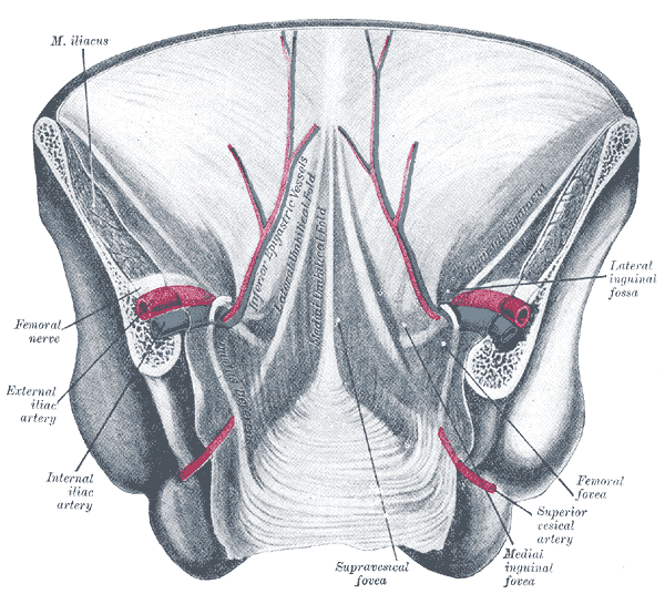
Caption = Posterior view of the anterior abdominal wall in its lower half. The peritoneum is in place, and the various cords are shining through.

Caption2 = The peritoneum of the male pelvis.
System =
Precursor =
MeshName =
MeshNumber =
DorlandsPre = l_09
DorlandsSuf = 12493498
The lateral umbilical fold overlies the inferior epigastric artery (a branch of the external iliac artery) and its accompanying veins. Unlike the median and medial umbilical folds, the contents of the Lateral Umbilical fold remain functional after birth.
Clinical significance
The lateral umbilical fold is an important reference site with regards to hernia classification. A direct hernia occurs medial to the lateral umbilical fold, whereas an indirect hernia originates lateral to the fold. This later case is due to the placement of the opening of the deep inguinal ring in the space lateral to the lateral umbilical fold, which allows the passage of the ductus deferens, testicular artery, and other components of the spermatic cord in men, or the round ligament of the uterus in women.
ee also
* Median umbilical ligament (some sources consider this the same structure)
* Medial umbilical ligament
External links
* Lateral umbilical fold
** SUNYAnatomyFigs|36|03|09 - "Internal surface of the anterior abdominal wall."
** SUNYAnatomyImage|7|3|84
=Additional
Wikimedia Foundation.
2010.
Look at other dictionaries:
lateral umbilical ligament — n MEDIAL UMBILICAL LIGAMENT … Medical dictionary
Medial umbilical ligament — The peritoneum of the male pelvis. (Medial umbilical ligament labeled at bottom left.) … Wikipedia
Median umbilical ligament — Posterior view of the anterior abdominal wall in its lower half. The peritoneum is in place, and the various cords are shining through. Median umbilical ligament isn t labeled, but it is located just underneath the median umbilical fold, seen in… … Wikipedia
medial umbilical ligament — n a fibrous cord sheathed in peritoneum and extending from the pelvis to the navel that is a remnant of part of the umbilical artery in the fetus called also lateral umbilical ligament … Medical dictionary
umbilical ligament lateral — former name for the medial umbilical ligament, still sometimes used although incorrect and confusing; alternatively, the term is sometimes incorrectly used to denote the lateral umbilical fold … Medical dictionary
Ligament — Diagram of the right knee. Typical joint In … Wikipedia
Umbilical folds — Posterior view of the anterior abdominal wall in its lower half. Umbilical folds labeled near middle … Wikipedia
Ligament — A ligament is a tough band of connective tissue that connects various structures such as two bones. Ligament is a fitting term; it comes from the Latin ligare meaning to bind or tie. * * * 1. A band or sheet of fibrous tissue connecting two or… … Medical dictionary
Umbilical artery — Artery: Umbilical artery Fetal circulation; the umbilical vein is the large, red vessel at the far left. The umbilical arteries are purple and wrap around the umbilical vein … Wikipedia
Ovarian ligament — Ligament: Ovarian ligament Uterus and right broad ligament, seen from behind. The broad ligament has been spread out and the ovary drawn downward. The ligament of ovary is labeled at the center top. The suspensory ligament of the ovary (not… … Wikipedia