- Posterior interosseous artery
-
Artery: Posterior interosseous artery 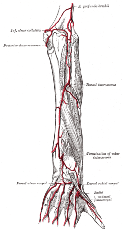
Arteries of the back of the forearm and hand. (Dorsal interosseus labeled at center right.) 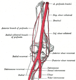
The Supinator. (Dorsal interosseus art. labeled at center right.) Latin arteria interossea posterior Gray's subject #152 596 Supplies Extensor digiti minimi, Extensor pollicis brevis Source Ulnar artery The posterior interosseous artery (dorsal interosseous artery) is an artery of the forearm.
It passes backward between the oblique cord and the upper border of the interosseous membrane. It appears between the contiguous borders of the supinator and the abductor pollicis longus, and runs down the back of the forearm between the superficial and deep layers of muscles, to both of which it distributes branches.
Where it lies upon the abductor pollicis longus and the extensor pollicis brevis, it is accompanied by the dorsal interosseous nerve. At the lower part of the forearm it anastomoses with the termination of the volar interosseous artery, and with the dorsal carpal network.
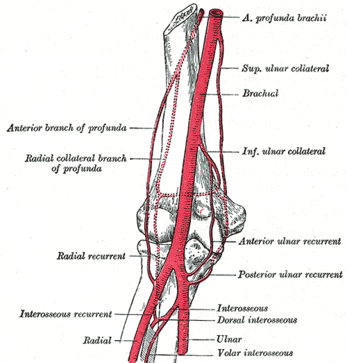
It gives off, near its origin, the interosseous recurrent artery, which ascends to the interval between the lateral epicondyle and olecranon, on or through the fibers of the Supinator, but beneath the anconeus, and anastomoses with the radial collateral branch of the profunda brachii, the posterior ulnar recurrent and the inferior ulnar collateral.
Additional images
-
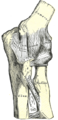
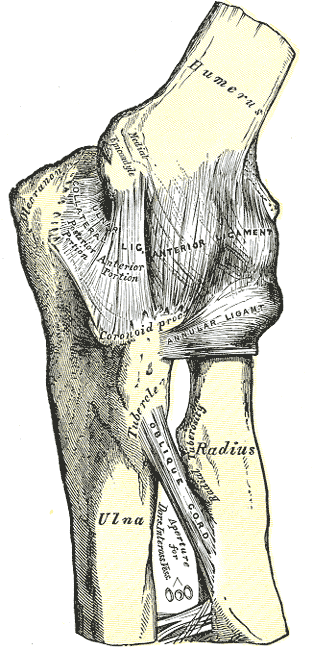
Left elbow-joint, showing anterior and ulnar collateral ligaments.
-

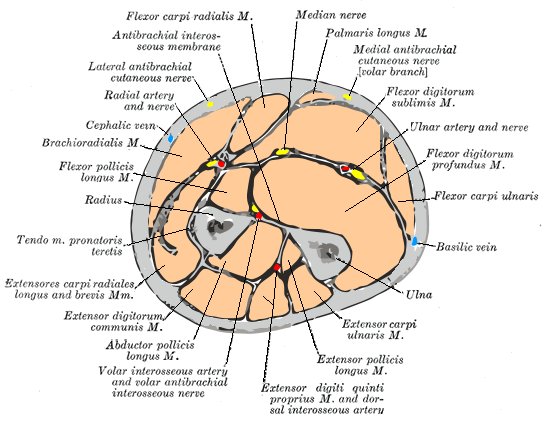
Cross-section through the middle of the forearm.
See also
External links
- EatonHand vas-026
- lesson4artofforearm at The Anatomy Lesson by Wesley Norman (Georgetown University)
- Atlas of anatomy at UMich hand_blood3 - "Dorsum of the hand, deep dissection, posterior view"
This article was originally based on an entry from a public domain edition of Gray's Anatomy. As such, some of the information contained within it may be outdated.
List of arteries of upper limbs (TA A12.2.09, GA 6.575) Axillary Shoulderscapular anastomosis · 1st part superior thoracic
2nd part thoracoacromial (deltoid branch) · lateral thoracic
3rd part subscapular (circumflex scapular, thoracodorsal) · anterior humeral circumflex · posterior humeral circumflexBefore splitforearm: radial recurrent
hand: superficial palmar branch · princeps pollicis · Radialis indices artery (radial of index finger)
wrist/carpus: dorsal carpal branch · palmar carpal branchforearm: ulnar recurrent (anterior, posterior) · common interosseous (anterior, posterior, recurrent)
hand: deep palmar branch
wrist/carpus: dorsal carpal branch · palmar carpal branchArchesCategories:- Arteries of the upper limb
- Cardiovascular system stubs
-

Wikimedia Foundation. 2010.