- Intravascular lymphoma
-
Intravascular lymphoma, also referred to as angiotropic large cell lymphoma,[1], intralymphatic lymphomatosis,[2] intravascular lymphomatosis,[2], and "Malignant angioendotheliomatosis"[1], is a condition that may present with variable symptoms, including various cutaneous morphologies (that are often very subtle and non-specific),[1]:742 and CNS symptoms.[3]
Most intravascular lymphomas are of the B-cell lineage.[4][5]
Contents
Diagnosis
Intravascular lymphoma is a condition that may present with non-specific symptoms and, thus, may be difficult to diagnose in a timely manner.[6]
Presentations may include non-specific cutaneous lesions,[1]:742 and/or CNS involvement.[3]
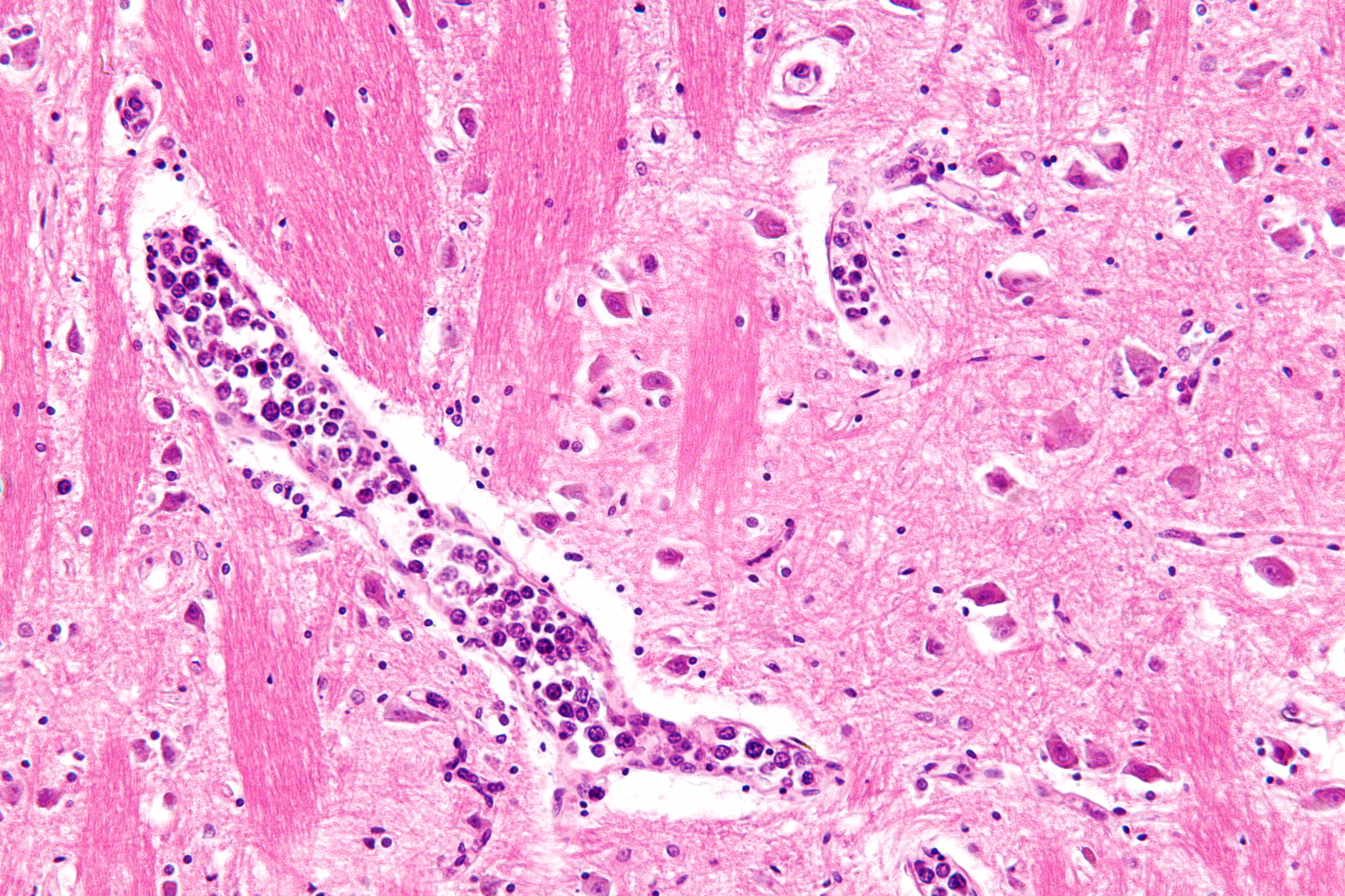
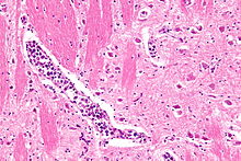
The diagnosis is established by the examination of tissue under the microscope (i.e. biopsy or autopsy). Intravascular lymphomas have a large cell morphology, i.e. the malignant cells are two or more times the size of a normal lymphocyte, and typically have a prominent nucleolus.
Classification
- Intravascular large B-cell lymphoma (ILBCL)
- Intravascular anaplastic large cell lymphoma
History
It used to be known as malignant angioendotheliomatosis, as it was once thought to arise from the endothelium.[7]
See also
- Proliferating angioendotheliomatosis
- List of cutaneous conditions
References
- ^ a b c d James, William D.; Berger, Timothy G.; et al. (2006). Andrews' Diseases of the Skin: clinical Dermatology. Saunders Elsevier. ISBN 0-7216-2921-0.
- ^ a b Rapini, Ronald P.; Bolognia, Jean L.; Jorizzo, Joseph L. (2007). Dermatology: 2-Volume Set. St. Louis: Mosby. ISBN 1-4160-2999-0.
- ^ a b Lapkuviene O, Forchetti D, Roepke JE (October 2001). "Unusual sites of involvement by hematologic malignancies. Case 1. Intravascular large B-cell lymphoma presenting with CNS symptoms". J. Clin. Oncol. 19 (19): 3988–91. PMID 11579120. http://jco.ascopubs.org/content/19/19/3988.full.
- ^ Wang L, Li C, Gao T (March 2010). "Cutaneous intravascular anaplastic large cell lymphoma". J Cutan Pathol 38 (2): 221–226. doi:10.1111/j.1600-0560.2010.01538.x. PMID 20337769.
- ^ Christopher D. M. Fletcher (5 April 2007). Diagnostic histopathology of tumors. Elsevier Health Sciences. pp. 1204–. ISBN 9780443074349. http://books.google.com/books?id=RniyoXeNmMwC&pg=PA1204. Retrieved 19 November 2010.
- ^ Ferry JA (April 2008). "Extranodal lymphoma". Arch. Pathol. Lab. Med. 132 (4): 565–78. doi:10.1043/1543-2165(2008)132[565:EL]2.0.CO;2. PMID 18384208.
- ^ Sheibani K, Battifora H, Winberg CD, et al. (April 1986). "Further evidence that "malignant angioendotheliomatosis" is an angiotropic large-cell lymphoma". N. Engl. J. Med. 314 (15): 943–8. doi:10.1056/NEJM198604103141502. PMID 3485768.

This cutaneous condition article is a stub. You can help Wikipedia by expanding it.